عفونت های شایع واژن

دکتر مریم رهنمون : عفونت های شایع واژن


در بانوان غدد داخل واژن و دهانه رحم به طور مداوم مقدار کمی مایع ترشح می کنند. این مکانیسم کمک به سلامت و پاک شدن مجرای تناسلی می کند. این ترشحات اکثراً بیرنگ و یا حداکثر شیری کم رنگ و بدون بو هستند، ولیکن بسته به سیکل ماهیانه در خانم ها رنگ و غلظت آن تغییر پذیر است. به طوریکه در زمان تخمک گذاری، حاملگی،شیر دادن و نزدیکی جنسی مقدار این ترشحات بیشتر می شود . بنابراین با توجه به نکات ذکر شده باید توجه داشت که افزایش ترشحات واژن در زنان در بسیاری موارد طبیعی است، به ویژه اینکه ترشحات باعث مرطوب شدن مجرای تناسلی زنان می شود،که این خود کمک بسیار در پیشگیری از بیماری گوناگون در این ناحیه می کند.


چه زمانی این افزایش ترشح طبیعی نیست؟
اگر افزایش ترشح باعث تغییر رنگ، بو ، التهاب ناحیه تناسلی، خارش، سوزش بشود غیر طبیعی خواهد بود به ویژه اینکه اگر بیمار در زمان عادت ماهیانه نباشد ولیکن ترشح خونی داشته باشد، حتماً باید به پزشک مراجعه کند.

چه چیز باعث این تغییرات می شود؟
هر عاملی که تغییر در محیط طبیعی واژن بدهد، و به عبارتی PH آن را تغییر بدهد، سبب افزایش ترشح غیر طبیعی از واژن می شود مانند: استفاده از بعضی صابونها، آنتی بیوتیکها، بیماری قند، حاملگی، عفونتها، و یا حتی پاک کردن مجرای تناسلی توسط اسپری های مخصوص ناحیه تناسلی و یا Vaginal douchمی توانند این مشگل را سبب شوند.

چگونه Vaginal Douching سبب عفونت می شود.؟
بسیاری از بانوان فکر می کنند این روش بسیار خوبی برای پاکیزه نگهداشتن واژن است. اگر چنانچه احساس بو در ناحیه تناسلی می کنید این بو اکثراً از قسمت خارجی دستگاه تناسلی است و شما می توانید با آب و صابون قسمت خارجی دستگاه تناسلی را تمیز نگاه دارید و همان حمام روزانه برای این پاکیزگی کافی است.
در حالیکه با گرفتن دوش واژینال و استفاده از مواد شیمیائی محیط طبیعی واژن را به هم می ریزید که این خود سبب عفونت می شود. از طرفی دیگر توسط Vaginal Douching (شستشوی واژن) باکتریهای موجود در واژن را به قسمت دهانه رحم خود و خود رحم روانه می کنید که این خود سبب عفونت در این نواحی خواهد شد.

چه عوامل دیگری سبب افزایش ترشح واژن می شوند؟
افزایش ترشح واژن به گونه غیر طبیعی به دلایل گوناگون خواهد بود که به طور کلی به نام Vaginitis نامیده می شوند.
1- عفونتهای قارچی واژن
درواژن زنان قارچ به مقدار کم به طور طبیعی زندگی می کند، محیط اسیدی واژن مانع از رشد این قارچها می شود. حال اگر به دلیلی از اسیدی بودن محیط واژن کاسته شود قارچها رشد بیشتر خواهند کرد و سبب عفونت قارچی در واژن می شوند. حاملگی، بعضی از آنتی بیوتیکها، مصرف قرصهای جلوگیری از حاملگی، بیماری قند، استفاده از کورتون ، زمان سیکل ماهیانه، روابط جنسی، نیز از عواملی هستند که باعث تغییر محیط طبیعی واژن و افزایش رشد قارچها و در نتیجه باعث عفونت در واژن می شوند.

علائم عفونت قارچی چیست؟
باید بگویم که این بیماری بسیار شایع است. بیمار احساس نارحتی در ناحیه تناسلی دارد که به صورت سوزش، خارش در دهانه واژن و در قسمت خارجی ناحیه تناسلی احساس می شود. ترشحات واژن به رنگ سفید و بسیار غلیظ در می آید که می توان آن را به شکل تکه های ریز پنیر مانند دید. بیمار احساس سوزش در زمان نزدیکی دارد. و همینطور احساس تورم در قسمت خارجی دستگاه تناسلی می کند.
ولیکن به یاد داشته باشید که اگر مکرراً و یا به گونه شدید بیماری مبتلا شوید که گاهی هم به درمان جواب نمی دهد و بلافاصله پس از درمان بیماری باز می گردد باید از نظر سایر بیماریهای سیستمیک به ویژه بیماری قند بررسی شوید.

چگونه درمان می شوید؟
درمان این بیماری بسیار ساده و معمولا با استفاده از کرمهای موضعی ضد قارچ مخصوص دستگاه تناسلی بانوان و یا گاهی هم اگر قادر به استفاده از کرم واژینال نباشید، با خوردن دارو درمان پذیر می باشد. گاهی توسط داروهائی که احتیاج به نسخه پزشک ندارد این بیماری به راحتی درمان پذیر است. ولیکن باز هم تکرار می کنم، چنانچه حامله هستید و یا به طور مکرر درگیر این بیماری می شوید، باید حتماً به پزشک خود مراجعه کنید که علت اصلی را جویا شوید.

چگونه از ابتلا مکرر به این بیماری پیشگیری کنیم؟
به نکات زیر توجه داشته باشید.
•حتماً از شورت های کتانی استفاده کنید.
• تا آنجا که ممکن است از پوشیدن شورت های پلاستیکی و یا بسیار تنگ و چسبان خودداری کنید.
از کاغذهای توالت حاوی مواد معطر و رنگی، Tampon، Pad های معطر و Bubble Bath و یا به طور کلی نشستن در وان حمام پرهیز کنید.
• اگر باز هم گرفتار عفونت مکرر قارچی واژن شدید به پزشک خود حتماً مراجعه کنید به دو دلیل نخست اینکه ممکن است علت بیماریهای سیستمیک بدن باشد و دو م گاهی لازم است که پزشک شما را برای مدت چند ماهی روی درمان بگذارد که از تکرار عفونت جلوگیری شود.

آیا همسر و یا دوست پسر شما باید درمان بشود؟
در این عفونت یا ر شما احتیاج به درمان ندارد و این بیماری اکثراً او را گرفتار نخواهد کرد مگر اینکه همسر شما مشکل دیگری ازنظر بیماری سیستمیک داشته باشد.

عفونت های میکروبی واژن
این عفونت Bac terial Vaginitis خوانده می شود.
محیط واژن حاوی نوعی باکتری است که به نام باکتری خوب واژن معروف است و همینطور حاوی باکتری دیگری است که به نام Anacrobes نامیده می شوند. گاهی بدون دلیل ویژه این نوع باکتری بیش از انداره رشد کرده و سبب بیماری می شوند.

چه علائمی دارند؟
گاهی بیمار اصلاً علائمی ندارد، گاهی این بیماری سبب افزایش ترشح واژن می شود که اکثراً آبکی است. یا گاهی رنگ زرد کمرنگ یا پررنگ تر دارد. گاهی بودار می شود که به اصطلاح Fishy Smell خوانده می شود که بیماران این بو را اکثراً پس از نزدیکی با همسر احساس می کنند، و گاهی بیمار احساس سوزش و خارش دارد.

چگونه تشخیص داده می شود؟
به پزشک مراجعه کنید. ترشح شما را در مطب آزمایش می کند و بلافاصله تشخیص داده خواهد شد.

آیا این نوع عفونت ازطریق نزدیکی منتقل می شود؟
اگر چه این بیماری در بانوانی که فعالیت جنسی دارند بیشتر دیده می شود ولیکن ارتباطی به نزدیکی ندارد و یار مقابل هم احتیاج به درمان ندارد. مگر اینکه بیمار مکرراً گرفتار این بیماری بشود.

چگونه درمان می شود؟
با تمام سادگی این بیماری احتیاج به درمان دارد چراکه اگر درمان نشود عفونت به سمت دهانه رحم، رحم و لوله ها می رود و سبب مشکلات بسیار و بدتری خواهد شد. به ویژه اگر اینکه زن حامله باشد. این بیماری را می شود به طور موضعی و یا خوراکی درمان کرد ولی درمان این بیماری حتماً احتیاج به مراجعه به دکتر و نسخه پزشک دارد .

عفونت شایع دیگر واژنTrichumoniasis است
عامل این بیماری یک نوع انگل به نام تریکومونا است که اکثراِ در اثر نزدیکی با فرد مبتلا ، بیمار درگیر آن خواهد شد. گاهی بیمار مدتها این بیماری را دارد بدون اینکه علائمی داشته باشد.
چه علائمی دارد؟ افزایش ترشح واژن، تغییر رنگ آن به صورت آبکی، یا زرد رنگ و یا گاهی سبز کمرنگ، بوی بد، خارش و سوزش، درد و سوزش در هنگام دفع ادرار از علائم آن است. اکثراً احتیاج به درمان خوراکی دارد. در این نوع گرفتاری همسر بیمار هم بهتر است که درمان بشود.
در اینجا باید یاد آور شوم که عفونتهای دیگر ناحیه تناسلی در ارتباط با رابطه جنسی ونزدیکی بیمار به آنها مبتلا می شود ازقبیل «کلامید یا»، سوزاک، هم می تواند سبب افزایش ترشح واژن بشود، ولیکن اینها عفونت هائی هستند که ابتدا دهانه رحم را گرفتار می کنند و گاهی تنها علامت آنها افزایش ترشح واژن است که در این نوع عفونتها زن و مرد هر دو باید درمان بشوند. ولیکن به خاطر داشته باشید که در این مقاله فقط سخن بر سر عفونت های شایع ناحیه واژن است نه سایر قسمتهای تناسلی

نکاتی که برای پیشگیری از عفونت واژن باید رعایت شود.
1- هرگز از Vaginal douche استفاده نکنید
2- از مواد عطری- اسپری عطری، Tamponsو همینطور از Pad های معطر در زمان خونریزی ماهیانه پرهیز کنید.
3- همیشه شورت های کتانی که زیاد هم تنگ و چسبان نباشند، بپوشید.
4- همیشه ناحیه تناسلی و مقعد را از جلو به عقب پاک کنید این روش تا آنجا که ممکن است از انتقال عفونت از ناحیه رکتوم به واژن جلوگیری خواهد شد.
5- گاهی این خارش و سوزش در ناحیه تناسلی ممکن است به دلیل آلرژی به مواد پاک کننده باشد در این صورت بهتر است که پودر لباسشوئی و کاغذ یا مایع نرم کننده پارچه را در موقع شستشو عوض کنید.
6- گاهی کاندوم یــــا «دیافراگم» و یا Sperm Killing Gel که برای پیشگیری از حاملگی مصرف می شود ممکن است سبب حساسیت و خارش و سوزش در ناحیه واژن بشود، در اینصورت بهتر است روش دیگری را جایگزین کنید.
7- پس از دوش روزانه ناحیه خارجی تناسلی را با حوله خوب خشک کنید

الایزا  

) الایزا                


امروزه الايزا بعنوان يكي از قدرتمندترين روشهاي آزمايشگاهي – تحقيقاتي در جهان مطرح مي‌باشد. اين روش از حساسيت و اختصاصيت بسيار بالايي برخوردار است تا بدانجا كه هم‌ اكنون روش اصلي در زمينه‌ هاي مختلف علوم حياتي از شناسايي و تشخيص ويروسها، باكتري‌ها و انگل‌هاي پروتوزوا و متازو گرفته تا شناسايي مقادير بسيار كم هورمونها و مولكولهاي حياتي در پزشكي باليني و آزمونهاي تحقيقاتي مي‌باشد .

چگونگي انجام آزمون الايزا :


پايه اساسي آزمون الايزا براساس واكنش آنتي‌بادي با آنتي‌ژن مي‌باشد. در آزمون‌هاي الايزا يك آنتي‌بادي اختصاصي با يك آنتي‌ژن مشخص واكنش داده و سپس با استفاده از يك آنتي‌بادي اتصال يافته با يك آنزيم بعنوان سيستم نشانگر، آزمون ادامه يافته و درنهايت با افزودن سوبستراي آنزيم و تبديل سوبسترا به محصول كه يك مادة رنگي مي‌باشد آزمون الايزا به پايان مي‌رسد طول موج رنگ بدست آمده كه نشانگر حضور يك آنتي‌بادي (و يا آنتي‌ژن) و نيز غلظت آن مي‌باشد توسط يك دستگاه اسپكتروفتومتر قرائت شده و ثبت مي‌گردد.

بعنوان مثال در يكي از رايج‌ترين روشهاي الايزا ( روش غير مستقيم) كه براي شناسايي حضور و يا عدم  حضور آنتي‌بادي  برعليه يك آنتي‌ژن موردنظر بكار مي‌رود ابتدا يك آنتي‌ژن برروي سطح جامد پوشش داده  مي‌شود سپس نمونة مجهول كه حضور و يا عدم حضور آنتي‌بادي برعليه اين آنتي‌ژن درون آن مورد بررسي قرار مي‌گيرد بر روي اين سطح جامد پوشش داده شده با آنتي‌ژن, افزوده مي‌شود.  پس از آن آنتي‌بادي ثانويه يا درحقيقت آنتي‌بادي برعليه اين آنتي‌بادي اوليه كه متصل به آنزيم نيز مي‌باشد افزوده مي‌شود و در نهايت سوبستراي آنزيم در اختيار آنزيم قرار داده مي‌شود و پس از گذشت مدت زمان لازم واكنش آنزيمي خاتمه داده شده و رنگ بدست آمده توسط دستگاه اسپكتروفتومتر قرائت مي‌شود 

 مراحل انجام يك آزمون الايزا به‌صورت زير مي‌باشد:

 1 ) جذب يك آنتي‌ژن يا آنتي‌بادي به سطوح جامد پلاستيكي كه اصطلاحا پوشش‌دهي ناميده مي‌شود.

 2 ) افزودن نمونه‌هاي مورد آزمايش

3 ) انكوباسيون واكنشگرها براي دراختياز قراردادن مدت زمان كافي براي انجام واكنش

4) جدا نمودن واكنشگرهاي متصل شده و واكنش داده از واكنشگرهاي آزاد و متصل نشده با استفاده از عمل شستشو

5) افزودن عوامل متصل شده با آنزيم

6) انكوباسيون واكنشگرها براي دراختيار قراردادن مدت زمان كافي براي انجام واكنش

7) جدا نمودن واكنشگرهاي متصل شده و واكنش داده از واكنشگرهاي آزاد و متصل نشده با استفاده عمل شستشو

8) افزودن سوبستراي آنزيم جهت تشخيص واكنش دهنده‌ها

9) انكوباسيون واكنشگرها براي دراختيار قراردادن مدت زمان كافي براي انجام واكنش

10) خاتمه دادن واكنش آنزيمي توسط متوقف كننده‌ها و قرائت دانسيتة نوري بدست آمده توسط دستگاه اسپكتروفتومتر


 مترجم:ایمان بهروزفر


استرپتولازین ها

                      ( AS0 (ANTI-STREPTOLYSIN O

 استرپتولازین ها آنزیم هایی  هستند که بوسیله بتا استرپتو کوک های همولیتیک تولید می شوند .استر پتو لایزین ها شامل  streptolysin-s(stable)  و streptolysin-o(oxygen labile)می باشد.

..انتی بادی هایی که علیه این آنزیم ها تولید می شوند در خون افراد آلوده شده پدیدار می گردد و از این آنتی بادی در جهت تشخیص این عفونت ها استفاده می شود.به این آنتی بادی ها ANTISTREPTOLYSIN Oو یاباختصار ASOمی گویند

باختصار ASOمی گویند

اهمیت ASO

این مار کر باکتریایی(انتی بادی) یکی از اولین مارکر های رماتیسم و تب مخملک بحساب می آید.

مقادیر

از آنجایی که این انتی بادی ها بعنوان واکنش تاخیری ازدیاد حساسیت به باکتری استرپتو کوکوس تولید میشود . چیزی بعنوان مقدار نرمال) (normal valueوجود ندارد.هر گونه وجود این انتی بادی ها در خون بمعنی اینست که فرد در معرض این باکتریها قرار گرفته است.ازآ نجایی که بیشتر افراد در معرض این باکتری ها قرار میگیرند،اما علئم بالینی را نشان نمی دهند،حضور ASOصرفا نشان دهنده یک بیماری خطر ناک نیست.

مقادیر قابل قبول که حاکی از عدم وجود علایم بالینی می باشد به قرار زیر است:

بزرگسالان : کمتر از   unit200

کودکان: کمتر ازunit  300

اين مقادیر هنگامي حائز اهميت مي باشند كه بسیار افزایش پیدا کنند.همچنین این مقادیر بایستی با علائم بالینی همراه باشند.

اندازه گیری

از روش های سرولوژی  همانند لاتکس آگلو تیانسیون و اسلاید آگلو تیناسیون  والایزا در جهت تعیین مقدار دقیق ASOمیشود.

میکانیسم عمل

این آنتی بادی ها که علیه باکتری های مذکور تولید میشود با بعضی از انتی ژن های انسانی(همانند کلاژن)  cross reactionمیدهد.بنابر این حمله به ماده زمینه سلولی (ماتریکس) ارگان های مختلف هانند  قلب ، مفاصل ، پوست، مغز و.... رخ می دهد.

            متر جم:ایمان بهروزفر

              منبع: WIKIPEDIA


اکنه ( جوش صورت و پوست ، غرور جوانی Acne vulgaris

درمان اکنه

خود درمانی اکنه با اسید سالی سیلیک موضعی


درمانی که در پیشگیری  از پیدایش « کومدوهای کوچک» کارآیی دارد در پیشگیری  از آسیب های التهابی نیز کارآیی خواهد داشت که کومدوهای کوچک، درون آنها امکان رشد دارند.


 اکنه ( جوش صورت و پوست ، غرور جوانی Acne vulgaris ) ناراحتی بسیار شایعی است و بیش از 80% جوانهای 13 تا 19 ساله و بالاتر را گرفتار می کند،اگر چه اکنه معمولاً نخستین بار در اواخر کودکی یا بین 13 تا 16 سال ظاهر می شود ولی در بعضی مواقع  در 20 یا 30 سالگی خودنمایی می کند. بیشترین موارد ظهور  اکنه که در حدود 17 سالگی است، در هر دو جنس تقریباً یکسان است ولی در مردان شدت بیشتری دارد. اگر چه اکنه بیماری خطرناکی نیست، ولی می تواند استرس عاطفی و مسایل  روانشناختی مثل کناره گیری از اجتماع و گریز از مدرسه به خصوص برای 13 تا 19 ساله ها به بار آورد.اکنه در بیشتر جوانهای  13 تا 19 ساله خفیف است، در کمتر از ثلث آنها نه چندان شدید و در 2% شدید است. بیشتر مبتلایان برای رفع ناراحتی خود به پزشک مراجعه نمی کنند. بیشتر افراد بالغ اکنه خود را با توصیه ی داروساز یا بدون آن – با داروی بی نسخه – درمان می کنند.  بیماریزایی اکنهاگر چه چند عامل در بیماریزایی اکنه دخالت دارند، ولی نخستین علت در بیشتر افراد بالغ افزایش تراز هورمون جنسی نر ( androgen)، همراه سن بلوغ است. در طول بلوغ، افزایش تراز هورمون جنسی نر غده ها چربی ضمیمه مو را تحریک می کنند و آنها چربی بیشتر ترشح می نمایند. جمع شدن چربی غده ی ضمیمه ی موها به دلیلی ناشناخته، غالباً باعث تغییر کنش بافت پوششی فولیکولی می شود و حاصلش به وجود آمدن مواد مانع بر سر راه منفذ خارجی فولیکول و جلوگیری از خروج چربی اضافه از فولیکول می شود. به وجود آمدن این مواد مانع، باعث تغییر الگوی کراتینی شدن داخل فولیکول مو و غده ی چربی پوست می گردد.ماده کراتینی درون فولیکول معمولاً سازمانی شل دارد و اضافه بر آن ذرات تیغه مانند در آن بیشتر است و فقط معدودی ذره کراتوهیالین به همراه دارد. ماده ی کراتینی بخش پایینی قیف مانند فولیکول در جریان به وجود آمدن کومدو متراکمتر و ذرات تیغه مانند کمتر می شوند و ذرات کراتوهیالین افزایش می یابند و بعضی از سلولهای فولیکولی صاحب ماده ی بی شکلی می شوند که احتمالاً  چربی است و در فرآیند به وجود آمدن کراتین تولید می گردند. این فرایند نخستین  ضایعه اکنه را به وجود می آورد که همان میکرو کومدو است. گفته شده است که میکروکومدو « بمب زمانی به موقع» اکنه و ضایعه ی پیشگام آن است که درمان مناسب عمدتاً متوجه آن است.به دنبال مراحل اولیه ی پیدایش ضایعه، چربی و کراتین محبوس شده، پشت مانع موجود در منفذ خارجی فولیکول، جمع می شوند و به صورت یک کومدوی بسته یا « سرسفید» در می آیند. در داخل کومدوی بسته است که باکتری بی هوازی Propionib actesium acne تکثیر می یابد و لیپازهایی تولید می کند که تری گلیسیریدهای چربی مو را به اسیدهای چرب تجزیه می نماید. این اسیدهای چرب، اگر فولیکول  را پاره کنند و داخل جلد نفوذ نمایند، موجب پیشرفت التهاب می گردند. میکرو کومدونهای پاره شده و کومدونهای بسته، با چنین فرایندی تبدیل به جوش، گرهک و کیست التهابی اکنه می شوند.رده بندی اکنهشایعترین رده بندی که مورد استفاده قرار دارد، از Pillsbury است که آنان را به چهار درجه تقسیم کرده است. در اکنه درجه یI کومدون عمده ترین ضایعه و جوشی است ملتهب  و کوچک. در آکنه ی درجه ی II ، وضع التهابی آشکارتر و تعداد جوشها و گرهکها بیشتر از آکنه درجه I است، ولی ضایعات عموماً  به صورت محدودند. در آکنه درجه ی III ، که آکنه ای بالنسبه سخت است، تعداد زیادی جوشهای کوچک و گاهی یک گرهک بزرگ دارد که عموماً  علاوه بر صورت در شانه ها و سینه ظاهر می شوند. آکنه ی درجه ی IV سخت ترین حالت آکنه است و معمولاً آن را acne conglobata می نامند.داروسازان عمدتاً با آکنه درجه I و II رو به رو می شوند. اکنه های سخت را باید به پزشک مراجعه کرد. پزشکان همه ی درجات آکنه را همراه دیگر بیماریها می بینند و دستورات خاص برای اکنه های درجه III و IV می دهند.  پیشگیری و درمانآکنه قابل درمان است ولی با موفقیت پیشگیری نمی شود. پاکیزگی عمومی، تغذیه ی خوب و ورزش بدنی منظم کمکی به پیشگیری است. درمانهای اولیه معمولاً از این نظر سودمند به نظر می رسند که از گسترش آکنه و سخت تر شدن آن پیشگیری می کند، از این گذشته درمان موثر آکنه نیز می تواند از پیدایش ضایعات جدید پیشگیری کند.درمان موضعی با داروهای بی نسخهدرمان عمده ی آکنه، بخصوص آکنه خفیف و نه چندان سخت، دارو درمانی موضعی است. بیشتر موارد آکنه کومدونی یا جوش مانند، می توانند با درمان سیستمی معالجه شوند. داروهای بی نسخه مثل « اسید سالی سیلیک» و « بنزویل پر اکسید»، گوگرد و « رزور سینول» سالها با موفقیت مورد استفاده بوده اند. FDA اسید سالی سیلیک 5/0 % تا 2% و رزورسینول 2%، را وقتی با گوگرد 3% تا 8% ، مخلوط  شود بی خطر و موثر برای داروهای بی نسخه در آدمی رده بندی کرده است ( گروه I). رده بندی بنزویل پراکسید در تابستان گذشته به وسیله FDA مورد تجدید ظنر قرار گرفته و از گروه I به گروه III انتقال داده شده است؛ زیرا در حیوانات بالقوه آغازگر تومور است. بررسی های بیشتری در حیوانات در دست انجامند تا معلوم گردد باید بار دیگر در گروه I جای داده شود یا نه.داروی موضعی برای آنکه موثر باشد نباید فقط ضایعه ی آکنه ی موجود را برطرف کند. بلکه باید از ظهور ضایعات جدید پیشگیری نماید. بنابراین برای به دست آوردن نتیجه ی مطلوب، داروها باید قابل کاربرد در تمامی ناحیه باشند نه حفظ  تک تک ضایعه ها. بهبود آکنه دست کم چند هفته طول می کشد. داروها کاملاً بی خطر انتخاب شده اند و تنها عارضه ی ناخواسته ی آنها سوزش موضعی است.اسید سالی سیلیکبیش از 100 سال است که اسید سالی سیلیک را برای درمان آکنه خفیف و نه چندان سخت به کار می برند. و پزشکان آزموده آن را درمانی موثر می شناسند که به خوبی تحمل می شود. اسید سالی سیلیک  جز فعال تعدادی از قلم های پاک کننده و مایعات  و کرم و ژل و لوسیون و بالشتک زخم و صابون است. کارآیی اسید سالی سیلیک در تعدادی از بررسیهای بالینی  و آزمایشهای رفع جوشهای کوچک  صورت نشان داده شده است.در فرآورده های دارای اسید سالی سیلیک ، مانند Stri Dex ، الکل  موجود به سرعت تبخیر می شود و اسید سالی سیلیک  باقی مانده را  روی پوست باقی می گذارد. این گونه فرآورده ها روی پوست دیده نمی شوند و بهتر از « بنزویل پراکسید»  تحمل می گردند. در یک بررسی کنترل شده ای که بیمار از ماهیت داروی به کار رفته آگاهی نداشت ( blinded) از فرآورده های اسید سالی سیلیک دار 5/0% و 1% و 2% استفاده و معلوم شد که در از بین بردن جوشهای کوچک صورت اثر چشمگیر دارند ولی فرآورده ی 2% ظاهراً در معکوس ساختن غیر عادی کراتینی  شدن فولیکول بهتر بود.درمانهای با نسخهپزشکانی که آکنه های سخت را درمان می کنند، ( داروهای قویتر موضعی مثل « ترتی نویین‌» و 10% «بنزویل پراکسید» نسخه می کنند. محلولهای موضعی حاوی آنتی بیوتیک، مثل اریترومایسین و کلیندامایسین نیز فراوان نسخه می شوند. درمان سیستمی شامل تتراسایکلین، اریترومایسین یا ماینوسایکلین است. گاهی « ایزوترتی نویین» برای بعضی از بیماران انتخابی دارای آکنه سخت نسخه می شود.آکنه خطر مرگ به همراه ندارد و عموماً خفیف یا نه چندان سخت است. با وجود این می تواند استرس عاطفی و حتی مسایل روانشناختی، بخصوص برای جوانان بین 13 تا 19 سال، به وجود آورد. این مسایل ممکن است تا سن کمال ادامه یابند و آکنه های سخت تر می توانند از طریق ایجاد عوارض پیجیده باقی گذاشتن اثر بر پوست ناراحتی ایجاد کنند. با وجود این اغلب آکنه ها با خوددرمانی و داروهایی که بر درمان و پیشگیری از ظهور ضایعات آکنه موثرند و بسیار خواستار دارد درمان می شوند.ازآنجا که نخستین ضایعه ی آکنه همان میکروکومدواست، درمانی که از ظهور آن جلوگیری می کند، بر تبدیل میکروکومدو به ضایعه ی التهابی آکنه و پیشرفت آن نیز موثر خواهد بود. اسید سالی سیلیک به عنوان یک عامل پوسته پوسته کردن پوست بدن، ریختن پوست سطحی را پیش می برد و این عمل بر سرعت از بین رفتن کومدونها و ضایعات التهابی می افزاید. محلول اسید سالی سیلیک 5/0% و 2% کومدونها را به خوبی از بین می برد و در پیشگیری از به وجود آمدن میکروکومدونها و پاک کردن ضایعات موجود موثر است.    

بعضی از داروهای بی نسخه آکنه که اسید سالی سیلیک دارندAveeno(S.C.Johnson)Clearasil (Richardson/Vicks Personal)Clear By Design (SmithKline Beecham Consumer)Fostex(Westwood/Squibb)Listerex(WarnerLambert)OxyClean (SmithKline Beecham Consumer)Oxy Nightwatch(SmithKline Beecham Consumer)Pernox(Westwood/Squibb)Propa P.H(Del)SalAc(GenDerm)StriDex(Glenbrook)


آرتريت‌ روماتوييد كودكان

آرتريت‌ روماتوييد كودكان
توضيح‌ كلي‌

آرتريت‌ روماتوييد كودكان عبارت‌ است‌ از يك‌ بيماري‌ التهابي‌ بافت‌ همبند، عمدتاً بافت‌ مفاصل‌، كه‌ در كودكان‌ رخ‌ مي‌دهد. امكان‌ دارد با آرتريت‌ ناشي‌ از بيماري‌ لايم‌ اشتباه‌ شود. مفاصلي‌ كه‌ درگير مي‌شوند معمولاً عبارتند از زانو، آرنج‌، مچ‌ پا، و گردن‌. التهاب‌ مي‌تواند عضلات‌ نزديك‌ مفصل‌، غضروف‌، و غشاهاي‌ پوشانندة‌ مفصل‌ را نيز متأثر سازد. اين‌ بيماري‌ بين‌ سنين‌ 5-2 سالگي‌ آغاز مي‌شود، و اغلب‌ تا جواني‌ بهبود مي‌يابد. اين‌ بيماري‌ در دختران‌ چهار برابر شايع‌تر از پسران‌ است‌.
                      
علايم‌ شايع‌

درد، تورم‌، و خشكي‌ در مفاصل‌ انگشتان‌ پا، زانو، مچ‌ پا، آرنج‌، شانه‌، يا گردن‌. درد ممكن‌ است‌ به‌ طور ناگهاني‌ يا تدريجي‌ آغاز شود و ممكن‌ است‌ در تنها يك‌، يا بسياري‌ از مفاصل‌ وجود داشته‌ باشد. امكان‌ دارد كودك‌ بدون‌ اين‌ كه‌ قادر به‌ توضيح‌ باشد، از راه‌رفتن‌ امتناع‌ كند.
دماي‌ بدن‌ هر روز و معمولاً در عصر تا حدود 4/39 درجة‌ سانتيگراد بالا مي‌رود. تب‌ به‌ طور شايع‌ با بثورات‌ پوستي‌ و لرز همراه‌ است‌.
بي‌اشتهايي‌، كاهش‌ وزن‌
كم‌خوني‌
تحريك‌پذيري‌، بي‌حالي‌
تورم‌ گره‌هاي‌ لنفاوي‌
درد و قرمزي‌ چشم‌
درد قفسة‌ صدري‌ به‌ قدري‌ شديد است‌ كه‌ بر قلب‌ تأثير مي‌گذارد.
                                                    
علل‌

احتمالاً به‌ علت‌ اختلال‌ خود ايمني‌ است‌ كه‌ طي‌ آن‌ دستگاه‌ ايمني‌ بدن‌ به‌ بافت‌ خودي‌ حمله‌ مي‌كند. بروز علايم‌ براي‌ اولين‌ بار، اغلب‌ باعث‌ استرس‌ جسماني‌ يا عاطفي‌ مي‌شود.

عوامل‌ افزايش‌ دهندة‌ خطر

ارث‌

پيشگيري‌

در حال‌ حاضر قابل‌ پيشگيري‌ نيست‌.

عواقب‌ مورد انتظار

اين‌ بيماري‌ در حال‌ حاضر غير قابل‌ درمان‌ است‌. اما در عين‌ حال‌، در 80%- 75% موارد، به‌ هنگام‌ بلوغ‌ يا اوايل‌ جواني‌، بيماري‌ به‌ كلي‌ فروكش‌ كرده‌ است‌.
هر حمله‌ معمولاً چند هفته‌ به‌ طول‌ مي‌انجامد و در طي‌ دوران‌ كودكي‌، بيماري‌ هر از چندگاهي‌ شعله‌ور مي‌شود و دوباره‌ فروكش‌ مي‌كند. علايم‌ را معمولاً مي‌توان‌ با درمان‌ تحت‌ كنترل‌ درآورد
عوارض‌ احتمالي‌

متأثر شدن‌ بافت‌هايي‌ غير از مفاصل‌، مثلاً التهاب‌ چشم‌، بزرگ‌ شدن‌ طحال‌، التهاب‌ پردة‌ دور قلب‌ يا عضلة‌ قلب‌.
تغيير شكل‌ و به‌ هم‌ خوردن‌ ساختمان‌ مفصل‌ به‌ طور دايمي‌
                                                  
درمان‌


اصول‌ كلي‌

آزمايش‌ خون‌، از جمله‌ بررسي‌ وضعيت‌ خود ايمني‌
عكس‌ برداري‌ از مفاصل‌. البته‌ تغييرات‌ مفصل‌ ممكن‌ است‌ تا مراحل‌ انتهايي‌ بيماري‌ نيز خود را در عكس‌ مفصل‌ نشان‌ ندهند.
روان‌ درماني‌ يا مشاوره‌ براي‌ كمك‌ به‌ خانوادة‌ كودك‌، تا آنها بتوانند به‌ خوبي‌ از عهدة‌ بيماري‌ طولاني‌ مدت‌ كودك‌ برآيند. شايد مهمترين‌ عامل‌ در درمان‌ كودك‌ حمايت‌ عاطفي‌ از وي‌ باشد.
جراحي‌ براي‌ درست‌كردن‌ مفصل‌ تغيير شكل‌ يافته‌ (گاهي‌)
اگر تشك‌ كودك‌ سفت‌ نيست‌، يك‌ تختة‌ ضخيم‌ چندلا زير تشك‌ قرار دهيد.
چشم‌ كودك‌ بايد حداقل‌ 2 بار در سال‌ معاينه‌ شود تا اگر التهابي‌ در چشم‌ وجود دارد زود تشخيص‌ داده‌ شود.
بسيار مهم‌ است‌ كه‌ كودك‌ به‌ مدرسه‌ عادي‌ به‌ طور روزانه‌ برود. هر جا كه‌ لازم‌ باشد، مدرسه‌ بايد خدمات‌ اضافي‌ براي‌ برآورده‌ كردن‌ نيازهاي‌ كودك‌ فراهم‌ كند.

داروها

آسپرين‌ يا ساير داروهاي‌ ضد التهاب‌ غير استروييدي‌، براي‌ كاهش‌ درد و التهاب‌
امكان‌ دارد داروهاي‌ ديگري‌ چون‌ كورتيكواستروييد، نمك‌ طلا، يا يا ساير داروها تجويز شوند

فعاليت‌

به‌ هنگام‌ حمله‌، كودك‌ بايد در تخت‌ استراحت‌ كند، مگر براي‌ توالت‌، تا زماني‌ كه‌ تب‌ و ساير علايم‌ تخفيف‌ يابند.
امكان‌ دارد براي‌ حفاظت‌ و حمايت‌ از مفصل‌ ملتهب‌ استفاده‌ از آتل‌ ضروري‌ باشد.
پس‌ از رفع‌ حمله‌، كودك‌ مي‌تواند به‌ تدريج‌ فعاليت‌هاي‌ عادي‌ را از سر بگيرد، البته‌ بايد در طي‌ روز ساعت‌هايي‌ را به‌ استراحت‌ بپردازد. كودك‌ نبايد زياد خسته‌ شود و هر شب‌ بايد حداقل‌ 12-10 ساعت‌ بخوابد.
فيزيوتراپي‌ توصيه‌ خواهد شد. بعضي‌ از حركات‌ را كودك‌ خود مي‌تواند انجام‌ دهد، و بعضي‌ از آنها را والدين‌ مي‌توانند براي‌ كودك‌ انجام‌ دهند. انجام‌ حركات‌ توصيه‌ شده‌ مهم‌ است‌ زيرا كمك‌ مي‌كند تا درد و اثرات‌ ناتوان‌ كنندة‌ بيماري‌ به‌ حداقل‌ برسند.
به‌ دليل‌ تغييرات‌ دوره‌اي‌ علايم‌، گاهي‌ برنامه‌ فيزيوتراپي‌ نياز به‌ بازبيني‌ خواهد داشت‌.
به‌ طور كلي‌ از انجام‌ ورزش‌هايي‌ كه‌ بدن‌ كودك‌ در معرض‌ ضربه‌ قرار مي‌گيرد بايد خودداري‌ شود، اما كودك‌ بايد تشويق‌ شود تا در ديگر فعاليت‌هاي‌ مدرسه‌، خانه‌ يا اجتماع‌ شركت‌ كند.

رژيم‌ غذايي‌

كودك‌ بايد يك‌ رژيم‌ مغذي‌ و متعادل‌ داشته‌ باشد، در عين‌ حال‌ وي‌ را بايد تشويق‌ به‌ خوردن‌ نمود.

در اين‌ شرايط‌ به‌ پزشك‌ خود مراجعه‌ نماييد

كودك‌ شما علايم‌ آرتريت‌ روماتوييد كودكان‌ را دارد.
علايم‌ زير به‌ هنگام‌ درمان‌ روي‌ دهند:
ـ درد قفسة‌ صدري‌ يا تب‌
ـ علايم‌ جديد و بدون‌ توجيه‌. توجه‌ داشته‌ باشيد كه‌ داروهاي‌ مورد استفاده‌ در درمان‌ ممكن‌ است‌ با عوارض‌ جانبي‌ همراه‌ باشند

منبع:خانه پزشکان ایران

صرع(epilepsy)

- صرع چیست؟

صرع را به زبان ساده می ‌توان به صورت زیر تعریف کرد: چنان چه تنظیم جریان الکتریکی فرد به هم بخورد، مدارهای کل مغز دچار اتصال شده و در نتیجه هوشیاری به هم می ‌خورد.

صرع فقط افتادن فرد و عدم هوشیاری نیست، بلکه به چندین شکل می ‌تواند دیده شود. به ‌طور مثال صرع می ‌تواند به صورت افت تحصیلی در مدرسه باشد، جا انداختن کلمات در جمله به خصوص در هنگام املا نوشتن اتفاق می‌ افتد و متاسفانه معلم‌ها فکر می ‌کنند که این دانش‌آموز دچار حواس پرتی می ‌باشد.


2- عوامل زمینه ‌ساز صرع کدامند؟

یکی از علل صرع به دوران قبل از تولد برمی ‌گردد، ولی به طور عمده علل صرع به بعد از تولد مربوط می‌ شود. هم چنین علل زیادی از انواع صرع ناشناخته می‌ باشد.

از علل قبل از تولد می ‌توان به عفونت‌های داخل رحمی (در جنین) اشاره کرد. از عفونت‌های شایع ایجاد کننده ی صرع در دوران بارداری سرخجه  و توکسو پلاسموز می ‌باشد. سایر عفونت‌های ویروسی، سوء تغذیه و مصرف الکل و دخانیات  در مادر هم می ‌تواند باعث صدمه به جنین شود.

از دیگر علل می ‌توان به نقص‌های ژنتیکی ایجاد شده در حین بارداری در جنین اشاره کرد که می ‌تواند عامل بروز تشنج در سال‌های آینده باشد. هم چنین قرار گرفتن در معرض محیط‌ های آلوده (مانند اشعه ایکس) می‌ تواند باعث آسیب به جنین شود.

از دلایل دیگر می ‌توان به ضربه‌ های مغزی جنین در حین بارداری یا موقع زایمان اشاره نمود.


دسته ی دیگر از دلایل صرع، مربوط به ضایعات در حین زایمان است که به عنوان فلج‌های مغزی عنوان می ‌شود. چنان چه در حین زایمان سر بچه آسیب ببیند و منجر به اختلالات انعقادی یا خونریزی‌های مغزی شود، می ‌تواند فرم‌هایی از تشنج را ایجاد کند. این آسیب‌ها هنگام تولد یا هفته ی اول بعد از تولد گاهی از علل بسیار مهم، برای تشنج‌های بعدی می ‌باشد.

در دوران نوزادی عفونت‌های مغزی مانند مننژیت  و آنسفالیت (تورم مغز در اثر ویروس‌ها)، و ضربه‌هایی که به خاطر جنب و جوش یا تصادف با ماشین به مغز وارد می شود، ممکن است باعث تشنج شود.

صرع و تشنج طوری نیست که بلافاصله بعد از دیدن ضربه، این بیماری ایجاد شود، بلکه ممکن است 10 تا 20 سال بعد خود را نشان بدهد و آسیب‌های در حین زایمان یا بعد از زایمان نتایج خود را در اواخر کودکی و یا بزرگسالی نشان دهد.

هم ‌چنین سوء تغذیه در زمان کودکی می ‌تواند یکی از دلایل تشنج در سنین بالاتر باشد، به خصوص کمبود مواد معدنی مثل کلسیم و منیزیم می ‌تواند از عوامل ایجاد کننده ی تشنج باشد.

در سنین بالا (افراد مسن) سکته ی مغزی  می ‌تواند باعث تشنج باشد و هم چنین به هم خوردن پایداری الکتریکی بدن باعث ایجاد صرع و تشنج در بیمار می ‌شود.

در افراد دیابتی  که از داروهای دیابتی برای پایین آوردن قند خون استفاده می ‌کنند یا بیمارانی که قند خون پایینی دارند، استعداد ابتلا به تشنج بیشتر از سایر افراد است.

با وجود تمام دلایل ذکر شده، علل درصد زیادی از انواع صرع در حال حاضر شناخته شده نمی ‌باشد.

آقای دکتر عسگر قربانی، متخصص بیماری‌های مغز و اعصاب(استادیار دانشگاه علوم پزشکی تهران) پاسخگوی سئوالات زیر می باشد.


1- راه‌های درمان تشنج و صرع کدام است؟

برای آن که بیماری صرع اثبات شود، باید ابتدا یک سری بیماری‌های مشابه شناخته شوند. یکی از بیماری‌های مشابه سنکوب می‌ باشد که خون به مقدار کافی به مغز نمی ‌رسد و به صورت گذرا در اثر دیدن مثلاً یک حادثه یا تصادف؛ یا در اثر گرمای یا سرمای زیاد، خون برای مدت کوتاهی به مغز نمی ‌رسد؛ یا به دنبال افت فشار به علت از دست دادن خون و شوک عصبی ممکن است فرد دچار سنکوب شود و بیافتد. این بیماران باید با توجه به شرح حال و معاینه، از بیماران صرعی افتراق داده شوند.

بعضی موارد در بیماری‌های قلبی یا سکته‌های مغزی  که خون کافی به مغز نمی ‌رسد ممکن است کاهش هوشیاری رخ دهد که نباید با صرع اشتباه شود.

بیمارهای دیگری مانند میگرن  ممکن است باعث از دست رفتن هوشیاری شود.

هم چنین کمبود مواد معدنی و تغذیه‌ای می ‌تواند سبب از دست دادن هوشیاری شود.

بعضی از ضایعات مغزی مثل عفونت‌های مغزی- تومورهای مغزی، فشار بالای جمجمه ممکن است بدون ایجاد تشنج باعث افت هوشیاری شوند، ولی گاهی اوقات این موارد خود باعث تشنج می ‌شوند.

از نوار مغز، آزمایشات اختصاصی خون، آزمایشات عمومی خون، CTS ، SCAN وMRI برای تأیید یا ردّ تشنج استفاده می ‌شود.

بعد از مشخص شدن تشنج، درمان به دو دسته ی دارویی و غیردارویی تقسیم می ‌شود.

درمان‌های غیردارویی بدین جهت دارای اهمیت زیادی می ‌باشند که شامل حذف عوامل خطرناک به خصوص برای پیش‌گیری از آسیب‌های مغزی می ‌باشد که از زمان جنینی شروع می ‌شود و شامل خودداری از مصرف دخانیات، عدم مصرف الکل، خودداری از محیط ‌های آلوده مثل رادیواکتیو می ‌باشد.

انجام ورزش و پیاده روی به ‌طور غیرمستقیم آمار تشنج‌ها را پایین می ‌آورد.

برای جلوگیری از تشنج باید فرد خواب منظم و کامل داشته باشد (بی ‌خوابی یکی از مهم ‌ترین عواملی است که باعث تشدید تشنج می ‌شود). باید به تغذیه هم ‌اهمیت زیادی داده شود و و لبنیات ، سبزیجات  و پروتئین  به مقدار کافی در برنامه ی غذایی بیمار گنجانده شود.

جلوگیری از چاقی و پرخوری و جلوگیری از استرس  و اضطراب  بسیار مهم می ‌باشد.

در درمان دارویی، با داروهای معمولی حدود 70- 60 درصد بیماران کنترل می‌ شوند و در درصد کمی از بیماران نیاز به درمان با چند دارو وجود دارد.

در مجموع حدود 85 درصد بیماران تشنجی با یک یا چند دارو کنترل می ‌شوند و در 20- 15درصد موارد ممکن است با مصرف دارو به خوبی کنترل نشوند یا احتمالا بهبود نیابند. این‌ها بیمارانی هستند که آسیب‌های شدید مغزی دارند (مانند بیماران عقب‌مانده ی ذهنی یا افراد دچار ناهنجاری‌های شدید مغزی مادرزادی). البته این بیماران نیز درمان می‌ شوند.

در مجموع با درمان کامل، حدود 95درصد بیماران طول عمر طبیعی دارند و ترس جامعه از این بیماری همیشه بی‌ مورد می ‌باشد.

البته درصد کمی ممکن است افراد وابسته‌ای باشند، ولی اکثر افراد با درمان دارویی یا غیردارویی در زندگی هیچ مشکلی را نخواهند داشت، چه بسا افراد به ظاهر سالم ممکن است در برخورد با مشکلات زندگی عاجز باشند ولی این افراد در زندگی خود هیچ مشکلی نداشته باشند.

داروهای ضد صرع را در خانم‌های باردار نیز می ‌توان استفاده کرد.


2- عدم درمان به موقع این بیماری چه عواقبی ممکن است داشته باشد؟

عدم درمان به موقع باعث می ‌شود که کانون ایجاد تشنج (کانون شورشی تشنج) به صورت فونکسیون و فیزیولوژیک دچار اختلال شود و اگر درمان نشود ممکن است باعث تغییرات ساختمانی در بافت مغز گردد و نواحی دیگر مغز را نیز درگیر کند و کارکرد فکری فرد را کم کند. بنابراین درمان این بیماران الزامی است.

درمان صرع، طولانی مدت و حدود دو تا سه سال می‌ باشد و بعد از این دوره درمان کامل می‌ شود.

درصد بسیار کمی از بیماران ممکن است درمان طولانی مدت داشته باشند.این افراد، افرادی هستند که در لب گیج‌گاهی اختلال دارند یا دارای آسیب‌های مغزی بارزی‌ هستند یا مشکل مادرزادی دارند، ولی به ‌طور غالب با درمان 3-2 ساله به طور کامل بهبود می یابند.


3- توصیه ی شما به این جوان 14 ساله چیست؟

یکی از شایع‌ترین انواعی که می ‌تواند ارثی هم باشد به نام "صرع پرشی" جوانان گفته می ‌شود که در واقع صبح‌ها و حتی حین صبحانه خوردن اتفاق می ‌افتد. این نوع تشنج با این که خوش ‌خیم است نیاز به درمان دارد و بیش‌تر با درمان طولانی مدت درمان می ‌شود. تشنج‌های خوش ‌خیم بر روی کارآیی فکری و اجتماعی تأثیری ندارند، ولی چون به هر حال این بیماری وجود دارد باید درمان شود.


4- توصیه ی کلی شما به این بیماران چیست؟

به طور کلی صرع بیماری درمان‌پذیر و خوش ‌خیم می ‌باشد و ترس جامعه از این بیماری کاملاً نا بجا و ناآگاهانه می ‌باشد و اکثر بیماران دچار صرع از خیلی از افراد جامعه بالاتر می باشند و تا آخر عمر ممکن است هیچ مشکلی نداشته باشند. آقایان و خانم‌های جوان مبتلا به بیماری صرع به‌ هیچ وجه نگران ازدواج و عواقب بعدی نباشند.

در مجموع چون یک بیماری خوش ‌خیم می ‌باشد، بهتر است در صورت ثابت شدن این بیماری توسط پزشک متخصص، اقدامات درمانی انجام گیرد و مطمئن باشید که درصد بسیار زیادی از این افراد (بالای 95درصد) به ‌طور کامل درمان خواهند شد.

وقتي يک فرد مصروع تصميم به ازدواج  مي گيرد، سؤالات متفاوتي براي وي وجود دارد. از جمله اين كه آيا مي تواند ازدواج كند يا خير؟ آيا بيماري او به فرزندش نيز به ارث مي رسد؟ و... ابتدا بايد به اين مسئله توجه داشته باشيم كه اگر يك فرد عادي با شرايط طبيعي بچه دار شود، احتمال اين كه فرزندش دچار صرع شود ، حدود نيم درصد است. حال اگر مادري مبتلا به صرع باشد، به دليل آنكه صرع ، طبيعتاً اساس ژنتيكي هم دارد، احتمال ابتلاي كودك به صرع، حدود 5 درصد است. علي رغم اين مسئله بايد در نظر داشت كه به احتمال 95 درصد ، مادر مصروع كودك سالمي را به دنيا مي آورد و اين شانس بسيار بالايي است.

به فردي كه مبتلا به صرع است ، توصيه مي شود قبل از ازدواج ، تحت مشاوره (خصوصاً

 مشاوره ژنتيك  ) قرار گيرد و در طي دوران بارداري حتماً تحت نظر پزشك زنان و متخصص مغز و اعصاب قرار گيرد تا بدين ترتيب هرگونه احتمال خطر براي كودك وي از بين برود. بدين ترتيب با بررسي صحيح و منطقي اين مسئله ، بيماران مصروع مي توانند يك زندگي و ازدواج موفق داشته و فرزندان سالمي به دنيا آورند.

برخي از انواع بيماري صرع اساس ژنتيك شناخته شده اي دارند (مانند صرع كوچك ) كه ما مي دانيم ممكن است به ارث برسند. ولي برخي ديگر از انواع آن اساس ژنتيك ندارند. مثلاً اگر فردي در اثر ضربه مغزي دچار بيمار صرع گردد، هيچ گونه علت ژنتيكي در اين مورد وجود ندارد. يا اگر كودكي در جريان زايمان دچار مشكلاتي از قبيل نرسيدن اكسيژن به مغز شود، احتمال ابتلاي وي به صرع در آينده وجود دارد. ولي چنانچه عوامل بيروني موجب به وجود آمدن اين اختلال باشد، اين بيماري به فرزند به ارث نمي رسد.

خوشبختانه در صرع، مواردي كه علل ژنتيك ندارند كم نيستند و در برخي از مواقع مي توان گفت انواع صرع اساس ژنتيك ندارد.

ضمناً توجه به اين نكته ضروري است كه علي رغم اينكه اين بيماري متأسفانه خوشنام نيست و افراد جامعه عقايد نادرستي در مورد آن دارند، ولي بايد ياد بگيريم كه آن را آگاهانه بشناسيم.

هر بيماري مزمن شرايط اجتماعي و عاطفي خاصي را ايجاد مي كند كه لازم است كيفيت زندگي در آن بررسي گردد.

صرع، اختلالي است كه در آن تخليه ناگهاني انرژي در مغز صورت مي گيرد و مي تواند با اختلالات حسي، حركتي و رفتاري توام گردد كه وقتي تخليه امواج به صورت عمومي باشد، موجب حذف هوشياري و شكل عمومي تشنج مي شود و وقتي هوشياري كاملاً از بين نرود به صورت تشنج موضعي، ساده يا پيچيده بروز مي كند.

در صرع به عنوان يك بيماري مزمن ، نمي توان حملات را پيش بيني كرد و هميشه دغدغه اين حملات با فرد مصروع وجود دارد. بر همين اساس عوامل مهمي در كيفيت زندگي مصروعين ( در كودكان و بزرگسالان) وجود دارد كه به آنها اشاره مي كنيم:


­ كيفيت مراقبت در كودكان

مهمترين مشكل در كودكان اين است كه افزايش يا كاهش بيش از حد مراقبت در آنان مي تواند بر كيفيت زندگي آنها تأثير بگذارد. اين مسئله به خصوص در مادران وجود دارد كه به علت علاقه شديد به فرزندشان مي توانند در اين مسئله سهم به سزايي داشته باشند.

-كيفيت آموزش

امكانات آموزشي در اين مورد بايد در مدرسه، خانواده و جامعه وجود داشته باشد. زيرا آموزش مناسب مي تواند در ادامه زندگي فرد تأثير به سزايي داشته باشد.

ازدواج مصروعين

فرد مصروع نبايد بيماري خود را در زمان ازدواج كتمان نمايد و بايد حقيقت را با صداقت به همسر آينده خود بگويد و با پزشك خود مشاوره اي را انجام دهد.

بارداري فرد مصروع

بهترين روش در بارداري اين است كه قبل از بارداري برنامه ريزي صحيحي در اين مورد وجود داشته باشد و فرد مصروع اين مسئله را با پزشك خود و پزشك زنان در ميان گذارد تا داروي مناسبي براي وي تجويز گردد و در طول دوران بارداري تحت نظر باشد تا اگر احساس شد مشكلي براي بيمار و جنين به وجود مي آيد با بررسي هاي انجام گرفته بتوان برخورد مناسبي با اين مسئله داشت.

داروها نيز در حين بارداري ادامه پيدا مي كند و نحوه زايمان هم بنا به تصميم پزشك است. بعد از زايمان نيز مادر مي تواند به فرزند خود شير دهد زيرا مصرف داروها تأثير منفي در فرزند وي ندارد، بلكه توجه به اين مسئله ضروري است كه اگر خانم باردار در دوران بارداري بدون مشاوره پزشك، خودسرانه داروي خود را قطع نمايد و دچار حمله تشنج گردد، اين امر مي تواند براي وي خطرناك باشد.


ازدواج مصروعين از ديدگاه روان پزشكي

روان پزشك نقش كليدي را در مورد مشكلات و بيماري هايي كه در كنار صرع وجود دارد، ايفا مي كند و مي تواند در مورد مداخلات دارويي ( تداخل عمل و اثر داروها با يكديگر ) جهت درمان بيماري هاي همراه با صرع ، راهنمايي و مشاوره به بيمار، همسر و خانواده او ارائه دهد.

يكي از مشكلات عمده، نگرش ها، دانش ها و باورهاي غلط فرد يا خانواده يا خانواده همسر او در مورد بيماري است. حتي علاوه بر آنها، اين باورهاي غلط در خود پزشكان نيز در مورد ازدواج يك فرد مصروع مي تواند وجود داشته باشد.

مسئله ديگر اين است كه بعضي از افراد ازدواج را نجات دهنده، شفا و راه حل قطعي مشكلات خود مي دانند و فكر مي كنند هر مشكل رواني-  جسمي كه وجود داشته باشد، ناشي از فشارها و مشكلات جنسي است و به دنبال آن حكم ازدواج را صادر مي كنند.

در زمان مشاوره روان پزشكي ، پزشك ابتدا به نگرش هاي فرد توجه مي نمايد. به اين كه آيا اين نگرش ها به ازدواج فرد صدمه مي زند؟ در غير اين صورت فرد را مجاز به ازدواج مي داند. دومين مسئله اي كه پزشك مد نظر قرار مي دهد، اين است كه آيا بيماري هاي همراه در يكي از طرفين وجود دارد يا خير؟ نكته ديگري كه در ازدواج بايد به آن توجه نمود كفايت رواني – عاطفي بيمار است و اين كه آيا فرد مصروعي كه حملات وي كنترل شده و مانعي براي ازدواج  ندارد، كفايت همسري، پدر يا مادر بودن، بارداري و … را دارد يا خير؟!

نكته بسيار كليدي و مهم در مشاوره قبل و بعد از ازدواج ، آموزش هاي جنسي است كه متأسفانه در كشور در مورد آن سكوت مي شود و حتي پدر و مادرها نيز نوعي خود سانسوري را در اين مورد پيش مي گيرند. لذا بهترين زمان اطلاعات گرفتن از بيماران، راجع به مسائل جنسي، زمان شروع بحث خواستگاري و ازدواج است كه بسياري از مراجعين در مورد اين موضوع يا اطلاعات ندارند يا اطلاعات غلطي دارند.

بنابراين كار كردن روي آموزش هاي جنسي، شناخت فرد روي فيزيولوژي خود، اطلاعات هورموني، اطلاعات آناتوميك، اطلاعات روان شناختي، دوران بارداري ، شناخت داروها، اثرات آن بر روي بارداري و جنين و… موضوعاتي هستند كه در قالب آنها مي توان اطلاعات مربوط به مسايل زناشويي، آموزش هاي جنسي و به ويژه جلوگيري از بارداري را به مراجعين ارائه كرد، چرا كه در ماه ها و سال هاي اوليه اين افراد بايد از بچه دار شدن جلوگيري نمايند و چگونگي تأثير اين داروها  بعد از ازدواج نيز بررسي گردد.

 منبع:tebyan

اكينوكوكوس گرانولوزوس

Echinococcus granulosus

اكينوكوكوس گرانولوزوس (Echinococcus granulosus )
نام بيماري: Echinococosis, Hydatid disease, Cystic Hydatosis, Hydatidosis disease

اين كرم يكي از آلودگيهاي جدي و مهم انسان به شمار رفته و در ايران نيز شايع حائز اهميت است. اين كرم از راسته سيكلوفيليده و خانواده تنيائيده بوده و سگ سانان ميزبان اصلي (نهايي) و علفخواران ميزبان واسط اصلي آن هستند و انسان بطور اتفاقي بدمرحله لاروي آن (به عنوان ميزبان واسط غيرفعال) مبتلا مي‏شوند.


تاكنون چهار گونه از جنس اكينوكوكوس شناسايي شده است كه در انسان قادر به ايجاد بيماريهاي  مختلفي هستند:

۱)E.granulosus (مسبب بيماري كيست هيداتيد)،

۲)E.multilocularis(مسبب كيست آلوئولي)

۳)E.vogeli(مسبب بيماري پلي كيستيك)و

۴)E.oligarthrus(مسبب بيماري پلي كيستيك).

اپيدميولوژي
جهان: بيماري تقريبا در تمامي‏نواحي گرمسيري و نيمه گرمسيري دنيا، خصوصا نواحي كه ارتباط نزديكي بين انسان و سگ وجود دارد، بوفور ديده ميشود.

ايران: هنوز آمار دقيقي در رابطه با ميزان شيوع الودگي افراد در ايران وجود ندارد. اما براساس مطالعات انجام شده برروي بيماران در بيمارستانها، برآورد مي‏شود از هر ۱۰۰ هزار نفر ۱۲/۱-۱ نفر مبتلا به هيداتيدوز باشند.

در رابطه با ميزان آلودگي در ميزبانهاي اصلي (خصوصا سگ) و ميزبانهاي واسط(خصوصا گوسفند، گاو و شتر) مطالعات نسبتا كاملي صورت گرفته است و در كل ميزان آلودگي سگها در استانهاي اصفهان، مازندران، آذربايجان غربي و ايلام بين ۴۵ تا ۶۳% و در ساير نواحي بطور متوسط ۲۰% بوده است(در سيستان و بلوچستان حدود ۳/۳%).

مورفولوژي
كرم: اين كرم باندازه بسيار كوچك (معمولا ۳-۶mm و گاهي تا ۹mm) بوده و داراي يك سر (اسكولكس)كروي شكل با چهار بادكش فنجاني شكل و يك روستلوم با ۲۸ تا ۵۰ قلاب(معمولا ۳۰ تا ۳۶)، يك گردن كوتاه، يك پروگلوتيد نارس، يك پروگلوتيد بالغ و يك (گاهي دو) پروگلوتيد بارور است طول عمر آن در سگ ۵ تا ۲۹ ماه است.

مرحله لاروي (Hydatid) : به مرحله لاروي (متاسستودي) ا.گرانولوزوس كه درداخل بدن ميزبان واسط (از جمله دام و انسان)تشكيل مي‏شود ، كيست هيداتيه گفته مي‏شود.

پس از خورده شدن تخم توسط ميزبان واسط جنين آن(انكوسفر) ازتخم خارج شده و از طريق گردش خون به بافت‏هاي مختلف رفته و در آنجا به فرم لاروي كرم كه به صورت حبابچه يا كيست است در مي‏آيد و ظرف ۳ ماه قطر آن به ۴-۵mm و ظرف ۵ ماه به ۱cm رسيده و از آن به بعد سالانه ۱cm(و گاهي تا ۵cm) به قطر آن اضافه مي‏شود.

اندازه كيست‏هاي هيداتيك بطور معمول ۵cm است ولي تا ۲۰cm هم گزارش شده است.

چرخه زندگي
كرمهاي بالغ در روده سگ و گاهي ساير سگ سانان( گربه، گرگ، شغال، روباه) كانگرو و ميمون زندگي مي‏كند. آخرين بند كرم محتوي تعداد زيادي تخم است كه در داخل روده ميزبان و يا در خارج تركيده و تخم‏ها آزاد مي‏شوند.

در پي خوردن سبزيجات و مواد غذايي آلوده، جدار تخم‏ها درمعده باز شده و جنين‏هاي كرم(انكوسفرها)آزاد شده و با نفوذ به جدار معده يا روده وارد جريان خون وريدي و لنفاتيك شده و در بافتهاي مختلف مستقر ميشوند و در آن مكان به فرم لاروي (كيست) تبديل مي‏شوند.

در صورت خورده شدن بافتهاي الوده به كيست توسط ميزبان قطعي، اسكولكس‏ها در معده و روده آزاد شده و ظرف ۶ تا ۹ هفته به كرم‏هاي بالغ بارور تبديل مي‏شوند.

بيماريزايي و تظاهرات باليني
انسان از طريق خوردن آب، سبزيجات، مواد غذايي و يا حتي ليس زدن سگ آلوده يا تماس دست با سگ و آلوده شدن دست و در نهايت خوردنتخم آلوده مي‏شود.

بيماري ايجاد شده در انسان به شدت آلودگي و محل تشكيل لارو(كيست) در بدن بستگي دارد.در كل، كبد(۶۰ تا ۷۰%)، ريه‏ها(۲۰ تا ۴۰%)، عضلات (۴%)، طحال(۳%)، بافتهاي نرم(۳%)، مغز(۳%)، استخوان(۲%) و ساير نواحي(۱%) محل‏هاي اصلي استقرار لاروها


Pseudomonas aeruginosa

Pseudomonas aeruginosa is member of the Gamma Proteobacteria class of Bacteria. It is a Gram-negative, aerobic rod belonging to the bacterial family Pseudomonadaceae. Since the revisionist taxonomy based on conserved macromolecules (e.g. 16S ribosomal RNA) the family includes only members of the genus Pseudomonas which are cleaved into eight groups. Pseudomonas aeruginosa is the type species of its group. which contains 12 other members.

Like other members of the genus, Pseudomonas aeruginosa is a free-living bacterium, commonly found in soil and water. However, it occurs regularly on the surfaces of plants and occasionally on the surfaces of animals. Members of the genus are well known to plant microbiologists because they are one of the few groups of bacteria that are true pathogens of plants.  In fact, Pseudomonas aeruginosa is occasionally a pathogen of plants. However, Pseudomonas aeruginosa has become increasingly recognized as an emerging opportunistic pathogen of clinical relevance. Several different epidemiological studies track its occurrence as a nosocomial pathogen and indicate that antibiotic resistance is increasing in clinical isolates.

Pseudomonas aeruginosa is an opportunistic pathogen, meaning that it exploits some break in the host defenses to initiate an infection.  In fact, Pseudomonas aeruginosa is the epitome of an opportunistic pathogen of humans. The bacterium almost never infects uncompromised tissues, yet there is hardly any tissue that it cannot infect if the tissue defenses are compromised in some manner. It causes urinary tract infections, respiratory system infections, dermatitis, soft tissue infections, bacteremia, bone and joint infections, gastrointestinal infections and a variety of systemic infections, particularly in patients with severe burns and in cancer and AIDS patients who are immunosuppressed. Pseudomonas aeruginosa infection is a serious problem in patients hospitalized with cancer, cystic fibrosis, and burns. The case fatality rate in these patients is near 50 percent.

Pseudomonas aeruginosa is primarily a nosocomial pathogen. According to the CDC, the overall incidence of P. aeruginosa infections in U.S. hospitals averages about 0.4 percent (4 per 1000 discharges), and the bacterium is the fourth most commonly-isolated nosocomial pathogen accounting for 10.1 percent of all hospital-acquired infections.

Characteristics


Pseudomonas aeruginosa
is a Gram-negative rod measuring 0.5 to 0.8 µm by 1.5 to 3.0 µm. Almost all strains are motile by means of a single polar flagellum.

The bacterium is ubiquitous in soil and water, and on surfaces in contact with soil or water.  Its metabolism is respiratory and never fermentative, but it will grow in the absence of O2 if NO3 is available as a respiratory electron acceptor.

The typical Pseudomonas bacterium in nature might be found in a biofilm, attached to some surface or substrate, or in a planktonic form, as a unicellular organism, actively swimming by means of its flagellum. Pseudomonas is one of the most vigorous, fast-swimming bacteria seen in hay infusions and pond water samples.

In its natural habitat Pseudomonas aeruginosa is not particularly distinctive as a pseudomonad, but it does have a combination of physiological traits that are noteworthy and may relate to its pathogenesis.

Pseudomonas aeruginosa has very simple nutritional requirements. It is often observed "growing in distilled water", which is evidence of its minimal nutritional needs.  In the laboratory, the simplest medium for growth of Pseudomonas aeruginosa consists of acetate as a source of carbon and ammonium sulfate as a source of nitrogen.

P. aeruginosa possesses the metabolic versatility for which pseudomonads are so renowned. Organic growth factors are not required, and it can use more than seventy-five organic compounds for growth.

• Its optimum temperature for growth is 37 degrees, and it is able to grow at temperatures as high as 42 degrees.

• It is tolerant to a wide variety of physical conditions, including temperature.  It is resistant to high concentrations of salts and dyes, weak antiseptics, and many commonly used antibiotics.
 
Pseudomonas aeruginosa has a predilection for growth in moist environments, which is probably a reflection of its natural existence in soil and water.

These natural properties of the bacterium undoubtedly contribute to its ecological success as an opportunistic pathogen. They also help explain the ubiquitous nature of the organism and its prominence as a nosocomial pathogen.

P. aeruginosa
isolates may produce three colony types. Natural isolates from soil or water typically produce a small, rough colony. Clinical samples, in general, yield one or another of two smooth colony types. One type has a fried-egg appearance which is large, smooth, with flat edges and an elevated appearance. Another type, frequently obtained from respiratory and urinary tract secretions, has a mucoid appearance, which is attributed to the production of alginate slime. The smooth and mucoid colonies are presumed to play a role in colonization and virulence.


Pseudomonas aeruginosa
colonies on agar


P. aeruginosa
strains produce two types of soluble pigments, the fluorescent pigment pyoverdin and the blue pigment pyocyanin. The latter is produced abundantly in media of low-iron content and functions in iron metabolism in the bacterium. Pyocyanin (from "pyocyaneus") refers to "blue pus", which is a characteristic of suppurative infections caused by Pseudomonas aeruginosa.


The soluble blue pigment pyocyanin is produced by many, but not all, strains of Pseudomonas aeruginosa

Pseudomonas aeruginosa
is notorious for its resistance to antibiotics and is, therefore, a particularly dangerous and dreaded pathogen. The bacterium is naturally resistant to many antibiotics due to the permeabiliity barrier afforded by its Gram-negative outer membrane. Also, its tendency to colonize surfaces in a biofilm form makes the cells impervious to therapeutic concentrations antibiotics. Since its natural habitat is the soil, living in association with the bacilli, actinomycetes and molds, it has developed resistance to a variety of their naturally-occuring antibiotics. Moreover, Pseudomonas maintains antibiotic resistance plasmids, both R-factors and RTFs, and it is able to transfer these genes by means of the bacterial mechanisms of horizontal gene transfer (HGT), mainly transduction and conjugation.
 
Only a few antibiotics are effective against Pseudomonas, including fluoroquinolones, gentamicin and imipenem, and even these antibiotics are not effective against all strains. The futility of treating Pseudomonas infections with antibiotics is most dramatically illustrated in cystic fibrosis patients, virtually all of whom eventually become infected with a strain that is so resistant that it cannot be treated.

Diagnosis

Diagnosis of P. aeruginosa infection depends upon isolation and laboratory identification of the bacterium. It grows well on most laboratory media and commonly is isolated on blood agar or eosin-methylthionine blue agar. It is identified on the basis of its Gram morphology, inability to ferment lactose, a positive oxidase reaction, its fruity odor, and its ability to grow at 42°C. Fluorescence under ultraviolet light is helpful in early identification of P. aeruginosa colonies. Fluorescence is also used to suggest the presence of P. aeruginosa in wounds.

Pathogenesis

For an opportunistic pathogen such as Pseudomonas aeruginosa, the disease process begins with some alteration or circumvention of normal host defenses. The pathogenesis of Pseudomonas infections is multifactorial, as suggested by the number and wide array of virulence determinants possessed by the bacterium. Multiple and diverse determinants of virulence are expected in the wide range of diseases caused, which include septicemia, urinary tract infections, pneumonia, chronic lung infections, endocarditis, dermatitis, and osteochondritis.

Most Pseudomonas infections are both invasive and toxinogenic. The ultimate Pseudomonas infection may be seen as composed of three distinct stages: (1) bacterial attachment and colonization; (2) local invasion; (3) disseminated systemic disease. However, the disease process may stop at any stage. Particular bacterial determinants of virulence mediate each of these stages and are ultimately responsible for the characteristic syndromes that accompany the disease.
 
Colonization

Although colonization usually precedes infections by Pseudomonas aeruginosa, the exact source and mode of transmission of the pathogen are often unclear because of its ubiquitous presence in the environment. It is sometimes present as part of the normal flora of humans, although the prevalence of colonization of healthy individuals outside the hospital is relatively low (estimates range from 0 to 24 percent depending on the anatomical locale).

The fimbriae of Pseudomonas will adhere to the epithelial cells of the upper respiratory tract and, by inference, to other epithelial cells as well. These adhesins appear to bind to specific galactose or mannose or sialic acid receptors on epithelial cells. Colonization of the respiratory tract by Pseudomonas requires fimbrial adherence and may be aided by production of a protease enzyme that degrades fibronectin in order to expose the underlying fimbrial receptors on the epithelial cell surface. Tissue injury may also play a role in colonization of the respiratory tract, since P. aeruginosa will adhere to tracheal epithelial cells of mice infected with influenza virus but not to normal tracheal epithelium. This has been called opportunistic adherence, and it may be an important step in Pseudomonas keratitis and urinary tract infections, as well as infections of the respiratory tract.

The receptor on tracheal epithelial cells for Pseudomonas pili is probably sialic acid (N-acetylneuraminic acid). Mucoid strains, which produce an exopolysaccharide (alginate), have an additional or alternative adhesin which attaches to the tracheobronchial mucin (N-acetylglucosamine). Besides pili and the mucoid polysaccharide, there are possibly  other cell surface adhesins utilized by Pseudomonas to colonize the respiratory epithelium or mucin. Also, it is possible that surface-bound exoenzyme S could serve as an adhesin for glycolipids on respiratory cells.

The mucoid exopolysaccharide produced by P. aeruginosa is a repeating polymer of mannuronic and glucuronic acid referred to as alginate. Alginate slime forms the matrix of the Pseudomonas biofilm which anchors the cells to their environment and in medical situations, it protects the bacteria from the host defenses such as lymphocytes, phagocytes, the ciliary action of the respiratory tract, antibodies and complement. Biofilm mucoid strains of P. aeruginosa are also less susceptible to antibiotics than their planktonic counterparts. Mucoid strains of P. aeruginosa are most often isolated from patients with cystic fibrosis and they are usually found in lung tissues from such individuals.

Invasion

The ability of Pseudomonas aeruginosa to invade tissues depends upon production of extracellular enzymes and toxins that break down physical barriers and damage host cells, as well as resistance to phagocytosis and the host immune defenses. As mentioned above, the bacterial capsule or slime layer effectively protects cells from opsonization by antibodies, complement deposition, and phagocyte engulfment.

Two extracellular proteases have been associated with virulence that exert their activity at the invasive stage: elastase and alkaline protease. Elastase has several activities that relate to virulence. The enzyme cleaves collagen, IgG, IgA, and complement. It also lyses fibronectin to expose receptors for bacterial attachment on the mucosa of the lung. Elastase disrupts the respiratory epithelium and interferes with ciliary function. Alkaline protease interferes with fibrin formation and will lyse fibrin. Together, elastase and alkaline protease destroy the ground substance of the cornea and other supporting structures composed of fibrin and elastin. Elastase and alkaline protease together are also reported to cause the inactivation of gamma interferon (IFN) and tumor necrosis factor (TNF).

P. aeruginosa
produces three other soluble proteins involved in invasion: a cytotoxin (mw 25 kDa) and two hemolysins. The cytotoxin is a pore-forming protein. It was originally named leukocidin because of its effect on neutrophils, but it appears to be cytotoxic for most eucaryotic cells. Of the two hemolysins, one is a phospholipase and the other is a lecithinase. They appear to act synergistically to break down lipids and lecithin. The cytotoxin and hemolysins contribute to invasion through their cytotoxic effects on neutrophils, lymphocytes and other eucaryotic cells.

One Pseudomonas pigment is probably a determinant of virulence for the pathogen. The blue pigment, pyocyanin, impairs the normal function of human nasal cilia, disrupts the respiratory epithelium, and exerts a proinflammatory effect on phagocytes. A derivative of pyocyanin, pyochelin, is a siderophore that is produced under low-iron conditions to sequester iron from the environment for growth of the pathogen. It could play a role in invasion if it extracts iron from the host to permit bacterial growth in a relatively iron-limited environment. No role in virulence is known for the fluorescent pigments.

Dissemination


Blood stream invasion and dissemination of Pseudomonas from local sites of infection is probably mediated by the same cell-associated and extracellular products responsible for the localized disease, although it is not entirely clear how the bacterium produces systemic illness. P. aeruginosa is resistant to phagocytosis and the serum bactericidal response due to its mucoid capsule and possibly LPS. The proteases inactivate complement, cleave IgG antibodies, and inactivate IFN, TNF and probably other cytokines. The Lipid A moiety of Pseudomonas LPS (endotoxin) mediates the usual pathologic aspects of Gram-negative septicemia, e.g. fever, hypotension, intravascular coagulation, etc. It is also assumed that Pseudomonas Exotoxin A exerts some pathologic activity during the dissemination stage (see below).

Toxinogenesis

P. aeruginosa
produces two extracellular protein toxins, Exoenzyme S and Exotoxin A. Exoenzyme S  has the characteristic subunit structure of the A-component of a bacterial toxin, and it has ADP-ribosylating activity (for a variety of eucaryotic proteins) characteristic of many bacterial exotoxins. Exoenzyme S is produced by bacteria growing in burned tissue and may be detected in the blood before the bacteria are. It has led to the suggestion that exoenzyme S may act to impair the function of phagocytic cells in the bloodstream and internal organs as a preparation for invasion by P. aeruginosa.

Exotoxin A
has exactly the same mechanism of action as the diphtheria toxin; it causes the ADP ribosylation of eucaryotic elongation factor 2 resulting in inhibition of protein synthesis in the affected cell. Although it is partially-identical to diphtheria toxin, it is antigenically-distinct. It utilizes a different receptor on host cells than diphtheria toxin, but otherwise it enters cells in the same manner and has the exact enzymatic mechanism. The production of Exotoxin A is regulated by exogenous iron, but the details of the regulatory process are distinctly different in C. diphtheriae and P. aeruginosa.

Exotoxin A appears to mediate both local and systemic disease processes caused by Pseudomonas aeruginosa. It has necrotizing activity at the site of bacterial colonization and is thereby thought to contribute to the colonization process. Toxinogenic strains cause a more virulent form of pneumonia than nontoxinogenic strains. In terms of its systemic role in virulence, purified Exotoxin A is highly lethal for animals including primates. Indirect evidence involving the role of exotoxin A in disease is seen in the increased chance of survival in patients with Pseudomonas septicemia that is correlated with the titer of anti-exotoxin A antibodies in the serum. Also, tox- mutants show a reduced virulence in some models.

Table 1 (below) is a summary of the virulence determinants of Pseudomonas aeruginosa. Table 2 is a brief description of the diseases caused by Pseudomonas aeruginosa.
 

Table 1. Summary of the Virulence Determinants of Pathogenic Pseudomonas aeruginosa
Adhesins
fimbriae (N-methyl-phenylalanine pili)
polysaccharide capsule (glycocalyx)
alginate slime (biofilm)

Invasins

elastase
alkaline protease
hemolysins (phospholipase and lecithinase)
cytotoxin (leukocidin)
siderophores and siderophore uptake systems
pyocyanin diffusible pigment

Motility/chemotaxis

flagella
Retractile pili?

Toxins

Exoenzyme S
Exotoxin A
Lipopolysaccharide

Antiphagocytic surface properties

capsules, slime layers
LPS
Biofilm construction

Defense against serum bactericidal reaction

slime layers, capsules, biofilm
LPS
protease enzymes

Defense against immune responses
capsules, slime layers, biofilm
protease enzymes

Genetic attributes
genetic exchange by transduction and conjugation
inherent (natural) drug resistance
R factors and drug resistance plasmids

Ecological criteria
adaptability to minimal nutritional requirements
metabolic diversity
widespread occurrence in a variety of habitats


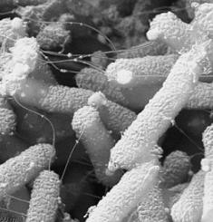
Pseudomonas aeruginosa Scanning electron micrograph. CDC


Table 2. Diseases caused by Pseudomonas aeruginosa

Endocarditis. Pseudomonas aeruginosa infects heart valves of IV drug users and prosthetic heart valves. The organism establishes itself on the endocardium by direct invasion from the blood stream.
 
Respiratory infections. Respiratory infections caused by Pseudomonas aeruginosa occur almost exclusively in individuals with a compromised lower respiratory tract or a compromised systemic defense mechanism. Primary pneumonia occurs in patients with chronic lung disease and congestive heart failure. Bacteremic pneumonia commonly occurs in neutropenic cancer patients undergoing chemotherapy. Lower respiratory tract colonization of cystic fibrosis patients by mucoid strains of Pseudomonas aeruginosa is common and difficult, if not impossible, to eradicate.

Bacteremia and septicemia. Pseudomonas aeruginosa causes bacteremia primarily in immunocompromised patients. Predisposing conditions include hematologic malignancies, immunodeficiency relating to AIDS, neutropenia, diabetes mellitus, and severe burns. Most Pseudomonas bacteremia is acquired in hospitals and nursing homes. Pseudomonas accounts for about 25 percent of all hospital acquired Gram-negative bacteremias.

Central nervous system infections. Pseudomonas aeruginosa causes meningitis and brain abscesses. The organism invades the CNS from a contiguous structure such as the inner ear or paranasal sinus, or is inoculated directly by means of head trauma, surgery or invasive diagnostic procedures, or spreads from a distant site of infection such as the urinary tract.

Ear infections including external otitis. Pseudomonas aeruginosa is the predominant bacterial pathogen in some cases of external otitis, including "swimmer's ear". The bacterium is infrequently found in the normal ear, but often inhabits the external auditory canal in association with injury, maceration, inflammation, or simply wet and humid conditions.

Eye infections. Pseudomonas aeruginosa can cause devastating infections in the human eye. It is one of the most common causes of bacterial keratitis, and has been isolated as the etiologic agent of neonatal ophthalmia. Pseudomonas can colonize the ocular epithelium by means of a fimbrial attachment to sialic acid receptors. If the defenses of the environment are compromised in any way, the bacterium can proliferate rapidly through the production of enzymes such as elastase, alkaline protease and exotoxin A, and cause a rapidly destructive infection that can lead to loss of the entire eye.

Bone and joint infections. Pseudomonas infections of bones and joints result from direct inoculation of the bacteria or the hematogenous spread of the bacteria from other primary sites of infection. Blood-borne infections are most often seen in IV drug users and in conjunction with urinary tract or pelvic infections. Pseudomonas aeruginosa has a particular tropism for fibrocartilagenous joints of the axial skeleton. Pseudomonas aeruginosa causes chronic contiguous osteomyelitis, usually resulting from direct inoculation of bone and is the most common pathogen implicated in osteochondritis after puncture wounds of the foot.

Urinary tract infections. Urinary tract infections (UTI) caused by Pseudomonas aeruginosa are usually hospital-acquired and related to urinary tract catheterization, instrumentation or surgery. Pseudomonas aeruginosa is the third leading cause of hospital-acquired UTIs, accounting for about 12 percent of all infections of this type. The bacterium appears to be among the most adherent of common urinary pathogens to the bladder uroepithelium. As in the case of E. coli, urinary tract infection can occur via an ascending or descending route. In addition, Pseudomonas can invade the bloodstream from the urinary tract, and this is the source of nearly 40 percent of Pseudomonas bacteremias.

Gastrointestinal infections. Pseudomonas aeruginosa can produce disease in any part of the gastrointestinal tract from the oropharynx to the rectum. As in other forms of Pseudomonas disease, those involving the GI tract occur primarily in immunocompromised individuals. The organism has been implicated in perirectal infections, pediatric diarrhea, typical gastroenteritis, and necrotizing enterocolitis. The GI tract is also an important portal of entry in Pseudomonas septicemia and bacteremia.

Skin and soft tissue infections, including wound infections, pyoderma and dermatitis. Pseudomonas aeruginosa can cause a variety of skin infections, both localized and diffuse. The common predisposing factors are breakdown of the integument which may result from burns, trauma or dermatitis; high moisture conditions such as those found in the ear of swimmers and the toe webs of athletes, hikers and combat troops, in the perineal region and under diapers of infants, and on the skin of whirlpool and hot tub users. Individuals with AIDS are easily infected. Pseudomonas has also been implicated in folliculitis and unmanageable forms of acne vulgaris.

Host Defenses


Most strains of P. aeruginosa  are resistant to killing in serum alone, but the addition of polymorphonuclear leukocytes results in bacterial killing. Killing is most efficient in the presence of type-specific opsonizing antibodies, directed primarily at the antigenic determinants of LPS. This suggests that phagocytosis is an important defense and that opsonizing antibody is the principal functional antibody in protecting from P. aeruginosa infections.
Once P. aeruginosa infection is established, other antibodies, such as antitoxin, may be important in controlling disease.

The observation that patients with diminished antibody responses (caused by underlying disease or associated therapy) have more frequent and more serious P. aeruginosa infections underscores the importance of antibody-mediated immunity in controlling Pseudomonas infections. unfortunately, cystic fibrosis is the exception. Most cystic fibrosis patients have high levels of circulating antibodies to bacterial antigens, but are unable to clear P. aeruginosa efficiently from their lungs. Cell-mediated immunity does not seem to play a major role in resistance or defense against Pseudomonas infections.

Epidemiology and Control of P. aeruginosa Infections

Pseudomonas aeruginosa
is a common inhabitant of soil, water, and vegetation. It is found on the skin of some healthy persons and has been isolated from the throat (5 percent) and stool (3 percent) of nonhospitalized patients. In some studies,  gastrointestinal carriage rates increased in hospitalized patients to 20 percent within 72 hours of admission.
Within the hospital, P. aeruginosa finds numerous reservoirs: disinfectants, respiratory equipment, food, sinks, taps, toilets, showers and mops. Furthermore, it is constantly reintroduced into the hospital environment on fruits, plants, vegetables, as well by visitors and patients transferred from other facilities. Spread occurs from patient to patient on the hands of hospital personnel, by direct patient contact with contaminated reservoirs, and by the ingestion of contaminated foods and water.

The spread of P. aeruginosa can best be controlled by observing proper isolation procedures, aseptic technique, and careful cleaning and monitoring of respirators, catheters, and other instruments. Topical therapy of burn wounds with antibacterial agents such as silver sulfadiazine, coupled with surgical debridement, dramatically reduces the incidence of P. aeruginosa sepsis in burn patients.

Pseudomonas aeruginosa is frequently resistant to many commonly used antibiotics. Although many strains are susceptible to gentamicin, tobramycin, colistin, and fluoroquinolins, resistant forms have developed. The combination of gentamicin and carbenicillin is frequently used to treat severe Pseudomonas infections. Several types of vaccines are being tested, but none is currently available for general use.

E. coli

Escherichia coli (E. coli) are members of a large group of bacterial germs that inhabit the intestinal tract of humans and other warm blooded animals (mammals, birds). Newborns have a sterile alimentary tract which within two days becomes colonized with E. coli

More than 700 serotypes of E. coli have been identified.  The different E. coli serotypes are distinguished by their “O” and “H” antigens on their bodies and flagella, respectively.  The E. coli serotypes that are responsible for the numerous reports of contaminated foods and beverages are those that produce Shiga toxin (Stx), so called because the toxin is virtually identical to that produced by another bacteria known as Shigella dysenteria type 1 (that also causes bloody diarrhea and hemolytic uremic syndrome [HUS] in emerging countries like Bangladesh) (Griffin & Tauxe, 1991, p. 60, 73).  The best known and most notorious Stx-producing E. coli is E. coli O157:H7.  It is important to remember that most kinds of E. coli bacteria do not cause disease in humans, indeed, some are beneficial, and some cause infections other than gastrointestinal infections, such urinary tract infections.  This section deals specifically with Stx-producing E. coli, including specifically E. coli O157:H7.

Shiga toxin is one of the most potent toxins known to man, so much so that the Centers for Disease Control and Prevention (CDC) lists it as a potential bioterrorist agent (CDC, n.d.).  It seems likely that DNA from Shiga toxin-producing Shigella bacteria was transferred by a bacteriophage (a virus that infects bacteria) to otherwise harmless E. coli bacteria, thereby providing them with the genetic material to produce Shiga toxin. 

Although E. coli O157:H7 is responsible for the majority of human illnesses attributed to E. coli, there are additional Stx-producing E. coli (e.g., E. coli O121:H19) that can also cause hemorrhagic colitis and post-diarrheal hemolytic uremic syndrome (D+HUS).  HUS is a syndrome that is defined by the trilogy of hemolytic anemia (destruction of red blood cells), thrombocytopenia (low platelet count), and acute kidney failure.

Stx-producing E. coli organisms have several characteristics that make them so dangerous.  They are hardy organisms that can survive several weeks on surfaces such as counter tops, and up to a year in some materials like compost.  They have a very low infectious dose meaning that only a relatively small number of bacteria (< 50) are needed “to set-up housekeeping” in a victim’s intestinal tract and cause infection.

The Centers for Disease Control and Prevention (CDC) estimates that every year at least 2000 Americans are hospitalized, and about 60 die as a direct result of E. coli infections and its complications. A recent study estimated the annual cost of E. coli O157:H7 illnesses to be $405 million (in 2003 dollars) which included $370 million for premature deaths, $30 million for medical care, and $5 million for lost productivity (Frenzen, Drake, and Angulo, 2005).

E. coli O157:H7—a foodborne pathogen

E. coli O157:H7 was first recognized as a foodborne pathogen in 1982 during an investigation into an outbreak of hemorrhagic colitis (bloody diarrhea) associated with consumption of contaminated hamburgers (Riley, et al., 1983).  The following year, Shiga toxin (Stx), produced by the then little-known E. coli O157:H7, was identified as the real culprit. 

In the ten years following the 1982 outbreak, approximately thirty E. coli O157:H7 outbreaks were recorded in the United States (Griffin & Tauxe, 1991). The actual number that occurred is probably much higher because E. coli O157:H7 infections did not become a reportable disease (required to be reported to public health authorities) until 1987 (Keene et al., 1991 p. 60, 73).  As a result, only the most geographically concentrated outbreaks would have garnered enough attention to prompt further investigation (Keene et al., 1991 p. 583).  It is important to note that only about 10% of infections occur in outbreaks, the rest are sporadic. 

The CDC has estimated that 85% of E. coli O157:H7 infections are foodborne in origin (Mead, et al., 1999).  In fact, consumption of any food or beverage that becomes contaminated by animal (especially cattle) manure can result in contracting the disease.  Foods that have been sources of contamination include ground beef, venison, sausages, dried (non-cooked) salami, unpasteurized milk and cheese, unpasteurized apple juice and cider (Cody, et al., 1999), orange juice, alfalfa and radish sprouts (Breuer, et al., 2001), lettuce, spinach, and water (Friedman, et al., 1999).

Escherichia coli

Escherichia coli

Theodor Escherich first described E. coli in 1885, as Bacterium coli commune, which he isolated from the feces of newborns. It was later renamed Escherichia coli, and for many years the bacterium was simply considered to be a commensal organism of the large intestine. It was not until 1935 that a strain of E. coli was shown to be the cause of an outbreak of diarrhea among infants.

The GI tract of most warm-blooded animals is colonized by E. coli within  hours or a few days after birth. The bacterium is ingested in foods or water or obtained directly from other individuals handling the infant. The human bowel is usually colonized within 40 hours of birth. E. coli can adhere to the mucus overlying the large intestine. Once established, an E. coli strain may persist for months or years. Resident strains shift over a long period (weeks to months), and more rapidly after enteric infection or antimicrobial chemotherapy that perturbs the normal flora. The basis for these shifts and the ecology of Escherichia coli in the intestine of humans are poorly understood despite the vast amount of information on almost every other aspect of the organism's existence. The entire DNA base sequence of the E. coli genome has been known since 1997.

E. coli is the head of the large bacterial family, Enterobacteriaceae, the enteric bacteria, which are facultatively anaerobic Gram-negative rods that live in the intestinal tracts of animals in health and disease. The Enterobacteriaceae are among the most important bacteria medically. A number of genera within the family are human intestinal pathogens (e.g. Salmonella, Shigella, Yersinia). Several others are normal colonists of the human gastrointestinal tract (e.g. Escherichia, Enterobacter, Klebsiella), but these bacteria, as well, may occasionally be associated with diseases of humans.

Physiologically, E. coli is versatile and well-adapted to its characteristic habitats. It can grow in media with glucose as the sole organic constituent. Wild-type E. coli has no growth factor requirements, and metabolically it can transform glucose into all of the macromolecular components that make up the cell. The bacterium can grow in the presence or absence of O2. Under anaerobic conditions it will grow by means of fermentation, producing characteristic "mixed acids and gas" as end products. However, it can also grow by means of anaerobic respiration, since it is able to utilize NO3, NO2 or fumarate as final electron acceptors for respiratory electron transport processes. In part, this adapts E. coli to its intestinal (anaerobic) and its extraintestinal (aerobic or anaerobic) habitats.

E. coli can respond to environmental signals such as chemicals, pH, temperature, osmolarity, etc., in a number of very remarkable ways considering it is a unicellular organism. For example, it can sense the presence or absence of chemicals and gases in its environment and swim towards or away from them. Or it can stop swimming and grow fimbriae that will specifically attach it to a cell or surface receptor. In response to change in temperature and osmolarity, it can vary the pore diameter of its outer membrane porins to accommodate larger molecules (nutrients) or to exclude inhibitory substances. With its complex mechanisms for regulation of metabolism the bacterium can survey the chemical contents in its environment in advance of synthesizing any enzymes that metabolize these compounds. It does not wastefully produce enzymes for degradation of carbon sources unless they are available, and it does not produce enzymes for synthesis of metabolites if they are available as nutrients in the environment.

E. coli is a consistent inhabitant of the human intestinal tract, and it is the predominant facultative organism in the human GI tract; however, it makes up a very small proportion of the total bacterial content. The anaerobic Bacteroides species in the bowel outnumber E. coli by at least 20:1. however, the regular presence of E. coli in the human intestine and feces has led to tracking the bacterium in nature as an indicator of fecal pollution and water contamination. As such, it is taken to mean that, wherever E. coli is found, there may be fecal contamination by intestinal parasites of humans.


Unstained cells of E. coli viewed by phase microscopy. about 1000X magnification. CDC.

Escherichia coli in the Gastrointestinal Tract

The commensal E. coli strains that inhabit the large intestine of all humans and warm-blooded animals comprise no more than 1% of the total  bacterial biomass.

The E. coli flora is apparently in constant flux. One study on the distribution of different E. coli strains colonizing the large intestine of women during a one year period (in a hospital setting) showed that 52.1% yielded one serotype, 34.9% yielded two, 4.4% yielded three, and 0.6% yielded four.  The most likely source of new serotypes of E. coli is acquisition by the oral route.

To study oral acquisition, the carriage rate of E. coli carrying antibiotic-resistance plasmids (R factors) was examined among vegetarians, babies, and nonvegetarians. It was assumed that nonvegetarians might carry more E. coli with R factors due to their presumed high incidence in animals treated with growth-promoting antimicrobial agents. However, omnivores had no higher an incidence of R-factor-containing E. coli than vegetarians, and babies had more resistant E. coli in their feces than nonvegetarians. No suitable explanation could be offered for these findings. Besides, investigation of the microbial flora of the uninhabited Krakatoa archipelago has shown the presence of antibiotic-resistant E. coli associated with plants.

The bottom line seems to be that most of us have more than one strain of E. coli in our gut, and intestinal strains tend to displace one another about three or four times a year.

Pathogenesis of E. coli

Over 700 antigenic types (serotypes) of E. coli are recognized based on O, H, and K antigens. At one time serotyping was important in distinguishing the small number of strains that actually cause disease. Thus, the serotype O157:H7 (O refers to somatic antigen; H refers to flagellar antigen) is uniquely responsible for  causing HUS (hemolytic uremic syndrome). Nowadays, particularly for diarrheagenic strains (those that cause diarrhea) pathogenic E. coli are classified based on their unique virulence factors and can only be identified by these traits. Hence, analysis for pathogenic E. coli usually requires that the isolates first be identified as E. coli before testing for virulence markers.

Pathogenic strains of E. coli are responsible for three types of infections in humans: urinary tract infections (UTI), neonatal meningitis, and intestinal diseases (gastroenteritis). The diseases caused (or not caused) by a particular strain of E. coli depend on distribution and expression of an array of virulence determinants, including adhesins, invasins, toxins, and abilities to withstand host defenses. These are summarized in Table 1 and applied to the discussion of pathogenic strains E. coli below.


Table 1. Summary of the Virulence Determinants of Pathogenic E. coli

Adhesins
CFAI/CFAII
Type 1 fimbriae
P fimbriae
S fimbriae
Intimin (non-fimbrial adhesin)
EPEC adherence factor

Invasins

hemolysin
Shigella-like "invasins" for intracellular invasion and spread

Motility/chemotaxis

flagella

Toxins
LT toxin
ST toxin
Shiga toxin
cytotoxins
endotoxin (LPS)

Antiphagocytic surface properties

capsules
K antigens
LPS

Defense against serum bactericidal reactions

LPS
K antigens

Defense against immune responses

capsules
K antigens
LPS
antigenic variation

Genetic attributes

genetic exchange by transduction and conjugation
transmissible plasmids
R factors and drug resistance plasmids
toxin and other virulence plasmids
siderophores and siderophore uptake systems
pathogenicity islands

Urinary Tract Infections

Uropathogenic E. coli (UPEC) cause 90% of the urinary tract infections (UTI) in anatomically-normal, unobstructed urinary tracts. The bacteria colonize from the feces or perineal region and ascend the urinary tract to the bladder. Bladder infections are 14-times more common in females than males by virtue of the shortened urethra. The typical patient with uncomplicated cystitis is a sexually-active female who was first colonized in the intestine with a uropathogenic E. coli strain. The organisms are propelled into the bladder from the periurethral region during sexual intercourse. With the aid of specific adhesins they are able to colonize the bladder.

The adhesin that has been most closely associated with uropathogenic E. coli is the P fimbria (or pyelonephritis-associated pili [PAP]). The letter designation is derived from the ability of P fimbriae to bind specifically to the P blood group antigen which contains a D-galactose-D-galactose residue. The fimbriae bind not only to red cells but to a specific galactose dissaccharide that is found on the surfaces uroepithelial cells in approximately 99% of the population.

The frequency of the distribution of this host cell receptor plays a role in susceptibility and explains why certain individuals have repeated UTI caused by E. coli. Uncomplicated E. coli UTI virtually never occurs in individuals lacking the receptors.

Uropathogenic strains of E. coli possess other determinants of virulence in addition to P fimbriae. E. coli with P fimbriae also possess the gene for Type 1 fimbriae, and there is evidence that P fimbriae are derived from Type 1 fimbriae by insertion of a new fimbrial tip protein to replace the mannose-binding domain of Type 1 fimbriae. In any case, Type 1 fimbriae could provide a supplementary mechanism of adherence or play a role in aggregating the bacteria to a specific manosyl-glycoprotein that occurs in urine.

Uropathogenic strains of E. coli usually produce siderophores that probably play an essential role in iron acquisition for the bacteria during or after colonization. They also produce hemolysins which are cytotoxic due to formation of transmembranous pores in host cell membranes. One strategy for obtaining iron and other nutrients for bacterial growth may involve the lysis of host cells to release these substances. The activity of hemolysins is not limited to red cells since the alpha-hemolysins of E. coli also lyse lymphocytes, and the beta-hemolysins inhibit phagocytosis and chemotaxis of neutrophils.

Another factor thought to be involved in the pathogenicity of the uropathogenic strains of E. coli is their resistance to the complement-dependent bactericidal effect of serum. The presence of K antigens is associated with upper urinary tract infections, and antibody to the K antigen has been shown to afford some degree of protection in experimental infections. The K antigens of E. coli are "capsular" antigens that may be composed of proteinaceous organelles associated with colonization (e.g., CFA antigens), or made of polysaccharides. Regardless of their chemistry, these capsules may be able to promote bacterial virulence by decreasing the ability of antibodies and/or complement to bind to the bacterial surface, and the ability of phagocytes to recognize and engulf the bacterial cells. The best studied K antigen, K-1, is composed of a polymer of N-acetyl neuraminic acid (sialic acid), which besides being antiphagocytic, has the additional property of being an antigenic disguise.
 

Neonatal Meningitis

Neonatal meningitis affects 1/2,000-4,000 infants. Eighty percent of E. coli strains involved synthesize K-1 capsular antigens (K-1 is only present 20-40% of the time in intestinal isolates).

E. coli strains invade the blood stream of infants from the nasopharynx or GI tract and are carried to the meninges.

The K-1 antigen is considered the major determinant of virulence among strains of E. coli that cause neonatal meningitis. K-1 is a homopolymer of sialic acid. It inhibits phagocytosis, complement, and responses from the host's immunological mechanisms. K-1 may not be the only determinant of virulence, however, as siderophore production and endotoxin are also likely to be involved.

Epidemiologic studies have shown that pregnancy is associated with increased rates of colonization by K-1 strains and that these strains become involved in the subsequent cases of meningitis in the newborn. Probably, the infant GI tract is the portal of entry into the bloodstream. Fortunately, although colonization is fairly common, invasion and the catastrophic sequelae are rare.

Neonatal meningitis requires antibiotic therapy that usually includes ampicillin and a third-generation cephalosporin.


Lysis of a dividing pair of E. coli cells in the presence of a beta-lactam antibiotic. Some beta lactam antibiotics, such as  ampicillin and cephalosporin, are effective in the treatment of meningitis caused by strains of E. coli (above). The beta lactam antibiotics prevent cell wall synthesis and assembly in the bacterium. When the bacterium grows in the presence of the antibiotic, the cell wall becomes progressively weaker and weaker, so the the organism eventually ruptures or "lyses", pouring out its cytoplasmic contents as shown here.

Intestinal Diseases Caused by E. coli

As a pathogen, E. coli is best known for its ability to cause intestinal diseases. Five classes (virotypes) of E. coli that cause diarrheal diseases are now recognized: enterotoxigenic E. coli (ETEC), enteroinvasive E. coli (EIEC), enterohemorrhagic E. coli (EHEC), enteropathogenic E. coli (EPEC), and enteroaggregative E. coli (EAEC). Each class falls within a serological subgroup and manifests distinct features in pathogenesis. A summary of the characteristics of diarrheagenic strains of E. coli is given in Table 2 at the end of this article.

Enterotoxigenic E. coli (ETEC)

ETEC is an important cause of diarrhea in infants and travelers in underdeveloped countries or regions of poor sanitation. In the U.S., it has been implicated in sporadic waterborne outbreaks, as well as due to the consumption of soft cheeses, Mexican-style foods and raw vegetables. The diseases vary from minor discomfort to a severe cholera-like syndrome. ETEC are acquired by ingestion of contaminated food and water, and adults in endemic areas evidently develop immunity. The disease requires colonization and elaboration of one or more enterotoxins. Both traits are plasmid-encoded.

ETEC may produce a heat-labile enterotoxin (LT) that is similar in molecular size, sequence, antigenicity, and function to the cholera toxin (Ctx). It is an 86kDa protein composed of an enzymatically active (A) subunit surrounded by 5 identical binding (B) subunits. It binds to the same identical ganglioside receptors that are recognized by the cholera toxin (i.e., GM1), and its enzymatic activity is identical to that of the cholera toxin.

ETEC may also produce a heat stable toxin (ST) that is of low molecular size and resistant to boiling for 30 minutes. There are several variants of ST, of which ST1a or STp is found in E. coli isolated from both humans and animals, while ST1b or STh is predominant in human isolates only. The ST enterotoxins are peptides of molecular weight about 4,000 daltons. Their small size explains why they are not inactivated by heat. ST causes an increase in cyclic GMP in host cell cytoplasm leading to the same effects as an increase in cAMP. ST1a is known to act by binding to a guanylate cyclase that is located on the apical membranes of host cells, thereby activating the enzyme. This leads to secretion of fluid and electrolytes resulting in diarrhea.

The infective dose of ETEC for adults has been estimated to be at least 108 cells; but the young, the elderly and the infirm may be susceptible to lower numbers.

ETEC adhesins are fimbriae which are species-specific. For example, the K-88 fimbrial Ag is found on strains from piglets; K-99 Ag is found on strains from calves and lambs; CFA I, and CFA II, are found on strains from humans. These fimbrial adhesins adhere to specific receptors on enterocytes of the proximal small intestine.

Symptoms ETEC infections include diarrhea without fever. The bacteria colonize the GI tract by means of a fimbrial adhesin, e.g. CFA I and CFA II, and are noninvasive, but produce either the LT or ST toxin. <>


Enteroinvasive E. coli (EIEC)

EIEC closely resemble Shigella in their pathogenic mechanisms and the kind of clinical illness they produce. EIEC penetrate and multiply within epithelial cells of the colon causing widespread cell destruction. The clinical syndrome is identical to Shigella dysentery and includes a dysentery-like diarrhea with fever. EIEC apparently lack fimbrial adhesins but do possess a specific adhesin that, as in Shigella, is thought to be an outer membrane protein. Also, like Shigella, EIEC are invasive organisms. They do not produce LT or ST toxin.

There are no known animal reservoirs of EIEC. Hence the primary source for EIEC appears to be infected humans. Although the infective dose of Shigella is low (in the range of 10 to few hundred cells), volunteer feeding studies showed that at least 106 EIEC organisms are required to cause illness in healthy adults. Unlike typical E. coli, EIEC are non-motile, do not decarboxylate lysine and do not ferment lactose. Pathogenicity of EIEC is primarily due to its ability to invade and destroy colonic tissue. The invasion phenotype, encoded by a high molecular weight plasmid, can be detected by PCR and probes for specific for invasion genes.


Enteropathogenic E. coli (EPEC)

EPEC induce a profuse watery, sometimes bloody, diarrhea. They are a leading cause of infantile diarrhea in developing countries. Outbreaks have been linked to the consumption of contaminated drinking water as well as some meat products.  Pathogenesis of EPEC involves a plasmid-encoded protein referred to as EPEC adherence factor (EAF) that enables localized adherence of bacteria to intestinal cells and a non fimbrial adhesin designated intimin, which is an outer membrane protein that mediates the final stages of adherence. They do not produce ST or LT toxins.

Adherence of EPEC strains to the intestinal mucosa is a very complicated process and produces dramatic effects in the ultrastructure of the cells resulting in rearrangements of actin in the vicinity of adherent bacteria. The phenomenon is sometimes called "attachment and effacing" of cells. EPEC strains are said to be "moderately-invasive",  meaning they are not as invasive as Shigella, and unlike ETEC or EAEC, they cause an inflammatory response. The diarrhea and other symptoms of EPEC infections probably are caused by bacterial invasion of host cells and interference with normal cellular signal transduction, rather than by production of toxins.

Through volunteer feeding studies the infectious dose of EPEC in healthy adults has been estimated to be 106 organisms.

Some types of EPEC are referred to as diffusely adherent E. coli (DAEC), based on specific patterns of adherence. They are an important cause of traveler's diarrhea in Mexico and in North Africa.

Enteroaggregative E. coli (EAEC)

The distinguishing feature of EAEC strains is their ability to attach to tissue culture cells in an aggregative manner. These strains are associated with persistent diarrhea in young children. They resemble ETEC strains in that the bacteria adhere to the intestinal mucosa and cause non-bloody diarrhea without invading or causing inflammation. This suggests that the organisms produce an enterotoxin of some sort. Recently, a distinctive heat-labile plasmid-encoded toxin has been isolated from these strains, called the EAST (EnteroAggregative ST) toxin. They also produce a hemolysin related to the hemolysin produced by E. coli strains involved in urinary tract infections. The role of the toxin and the hemolysin in virulence has not been proven. The significance of EAEC strains in human disease is controversial.

 
Enterohemorrhagic E. coli (EHEC)

EHEC are recognized as the primary cause of hemorrhagic colitis (HC) or bloody diarrhea, which can progress to the potentially fatal hemolytic uremic syndrome (HUS). EHEC are characterized by the production of verotoxin or Shiga toxins (Stx). Although Stx1 and Stx2 are most often implicated in human illness, several variants of Stx2 exist.

There are many serotypes of Stx-producing E. coli , but only those that have been clinically associated with HC are designated as EHEC. Of these, O157:H7 is the prototypic EHEC and most often implicated in illness worldwide. The infectious dose for O157:H7 is estimated to be 10 - 100 cells; but no information is available for other EHEC serotypes. EHEC infections are mostly food or water borne and have implicated undercooked ground beef, raw milk, cold sandwiches, water, unpasteurized apple juice and vegetables

EHEC are considered to be "moderately invasive". Nothing is known about the colonization antigens of EHEC but fimbriae are presumed to be involved. The bacteria do not invade mucosal cells as readily as Shigella, but EHEC strains produce a toxin that is virtually identical to the Shiga toxin. The toxin plays a role in the intense inflammatory response produced by EHEC strains and may explain the ability of EHEC strains to cause HUS. The toxin is phage encoded and its production is enhanced by iron deficiency.


E. coli O157:H7 Transmission EM. American Society for Microbiology

STAPHYLOCOCCUS

STAPHYLOCOCCUS

Clinically, the most important genus of the Micrococcaceae family is Staphylococcus. The Staphylococcus genus is classified into two major groups: aureus and non-aureus. S. aureus is a leading cause of soft tissue infections, as well as toxic shock syndrome (TSS) and scalded skin syndrome. It can be distinguished from other species of Staph by a positive result in a coagulase test(all other species are negative).

The pathogenic effects of Staph are mainly asssociated with the toxins it produces. Most of these toxins are produced in the stationary phase of the bacterial growth curve. In fact, it is not uncommon for an infected site to contain no viable Staph cells. The S. aureus enterotoxin causes quick onset food poisoning which can lead to cramps and severe vomiting. Infection can be traced to contaminated meats which have not been fully cooked. These microbes also secrete leukocidin, a toxin which destroys white blood cells and leads to the formation of pus and acne. Particularly, S. aureus has been found to be the causative agent in such ailments as pneumonia, meningitis, boils, arthritis, and osteomyelitis (chronic bone infection). Most S. aureus are penicillin resistant, but vancomycin and nafcillin are known to be effective against most strains.

Of the non-aureus species, S. epidermis is the most clinically significant. This bacterium is an opportunistic pathogen which is a normal resident of human skin. Those susceptible to infection by the bacterium are IV drug users, newborns, elderly, and those using catheters or other artificial appliances. Infection is easily treatable with vancomycin or rifampin.

    LABORATORY INDICATIONS:
  • Anaerobic glucose fermentation with acid production
  • Catalase +
  • Nitrate +
  • Coagulase +

Staphylococci

The Staphylococci

Staphylococci are Gram-positive spherical bacteria that occur in microscopic clusters resembling grapes. Bacteriological culture of the nose and skin of normal humans invariably yields staphylococci. In 1884, Rosenbach described the two pigmented colony types of staphylococci and proposed the appropriate nomenclature: Staphylococcus aureus (yellow) and Staphylococcus albus (white). The latter species is now named Staphylococcus epidermidis. Although more than 20 species of Staphylococcus are described in Bergey's Manual (2001), only Staphylococcus aureus and Staphylococcus epidermidis are significant in their interactions with humans. S. aureus colonizes mainly the nasal passages, but it may be found regularly in most other anatomical locales. S epidermidis is an inhabitant of the skin.

Taxonomically, the genus Staphylococcus is in the Bacterial family Staphylococcaceae, which includes three lesser known genera, Gamella, Macrococcus and Salinicoccus. The best-known of its nearby phylogenetic relatives are the members of the genus Bacillus in the family Bacillaceae, which is on the same level as the family Staphylococcaceae. The Listeriaceae are also a nearby family.

Staphylococcus aureus forms a fairly large yellow colony on rich medium, S. epidermidis has a relatively small white colony. S. aureus is often hemolytic on blood agar; S. epidermidis is non hemolytic. Staphylococci are facultative anaerobes that grow by aerobic respiration or by fermentation that yields principally lactic acid. The bacteria are catalase-positive and oxidase-negative. S. aureus can grow at a temperature range of 15 to 45 degrees and at NaCl concentrations as high as 15 percent. Nearly all strains of S. aureus produce the enzyme coagulase: nearly all strains of S. epidermidis lack this enzyme. S. aureus should always be considered a potential pathogen; most strains of S. epidermidis are nonpathogenic and may even play a protective role in their host as normal flora. Staphylococcus epidermidis may be a pathogen in the hospital environment.

Staphylococci are perfectly spherical cells about 1 micrometer in diameter. They grow in clusters because staphylococci divide in two planes. The configuration of the cocci helps to distinguish staphylococci from streptococci, which are slightly oblong cells that usually grow in chains (because they divide in one plane only). The catalase test is important in distinguishing streptococci (catalase-negative) from staphylococci, which are vigorous catalase-producers. The test is performed by adding 3% hydrogen peroxide to a colony on an agar plate or slant. Catalase-positive cultures produce O2 and bubble at once. The test should not be done on blood agar because blood itself contains catalase.


FIGURE 1. Gram stain of Staphylococcus aureus in pustular exudate
 

Table 1. Important phenotypic characteristics of Staphylococcus aureus


Gram-positive, cluster-forming coccus
nonmotile, nonsporeforming facultative anaerobe
fermentation of glucose produces mainly lactic acid
ferments mannitol (distinguishes from S. epidermidis)
catalase positive
coagulase positive
golden yellow colony on agar
normal flora of humans found on nasal passages, skin and mucous membranes
pathogen of humans, causes a wide range of suppurative infections, as well as food poisoning and toxic shock syndrome 

Pathogenesis of S. aureus infections

Staphylococcus aureus causes a variety of suppurative (pus-forming) infections and toxinoses in humans. It causes superficial skin lesions such as boils, styes and furunculosis; more serious infections such as pneumonia, mastitis, phlebitis, meningitis, and urinary tract infections; and deep-seated infections, such as osteomyelitis and endocarditis. S. aureus is a major cause of hospital acquired (nosocomial) infection of surgical wounds and infections associated with indwelling medical devices. S. aureus causes food poisoning by releasing enterotoxins into food, and toxic shock syndrome by release of superantigens into the blood stream.

S. aureus expresses many potential virulence factors: (1) surface proteins that promote colonization of host tissues; (2) invasins that promote bacterial spread in tissues (leukocidin, kinases, hyaluronidase); (3) surface factors that inhibit phagocytic engulfment (capsule, Protein A); (4) biochemical properties that enhance their survival in phagocytes (carotenoids, catalase production); (5) immunological disguises (Protein A, coagulase, clotting factor); and (6) membrane-damaging toxins that lyse eukaryotic cell membranes (hemolysins, leukotoxin, leukocidin; (7) exotoxins that damage host tissues or otherwise provoke symptoms of disease (SEA-G, TSST, ET (8) inherent and acquired resistance to antimicrobial agents.


FIGURE 2. Virulence determinants of Staphylococcus aureus
 

For the majority of diseases caused by S. aureus, pathogenesis is multifactorial, so it is difficult to determine precisely the role of any given factor. However, there are correlations between strains isolated from particular diseases and expression of particular virulence determinants, which suggests their role in a particular diseases. The application of molecular biology has led to advances in unraveling the pathogenesis of staphylococcal diseases. Genes encoding potential virulence factors have been cloned and sequenced, and many protein toxins have been purified. With some staphylococcal toxins, symptoms of a human disease can be reproduced in animals with the purified protein toxins, lending an understanding of their mechanism of action.

Human staphylococcal infections are frequent, but usually remain localized at the portal of entry by the normal host defenses. The portal may be a hair follicle, but usually it is a break in the skin which may be a minute needle-stick or a surgical wound. Foreign bodies, including sutures, are readily colonized by staphylococci, which may makes infections difficult to control. Another portal of entry is the respiratory tract. Staphylococcal pneumonia is a frequent complication of influenza. The localized host response to staphylococcal infection is inflammation, characterized by an elevated temperature at the site, swelling, the accumulation of pus, and necrosis of tissue. Around the inflamed area, a fibrin clot may form, walling off the bacteria and leukocytes as a characteristic pus-filled boil or abscess. More serious infections of the skin may occur, such as furuncles or impetigo. Localized infection of the bone is called osteomyelitis. Serious consequences of staphylococcal infections occur when the bacteria invade the blood stream. A resulting septicemia may be rapidly fatal; a bacteremia may result in seeding other internal abscesses, other skin lesions, or infections in the lung, kidney, heart, skeletal muscle or meninges.


FIGURE 3. Sites of infection and diseases caused by Staphylococcus aureus
 

Adherence to Host Cell Proteins

S. aureus cells express on their surface proteins that promote attachment to host proteins such as laminin and fibronectin that form the extracellular matrix of epithelial and endothelial surfaces. In addition, most strains express a fibrin/fibrinogen binding protein (clumping factor) which promotes attachment to blood clots and traumatized tissue. Most strains of S. aureus express both fibronectin and fibrinogen-binding proteins. In addition, an adhesin that promotes attachment to collagen has been found in strains that cause osteomyelitis and septic arthritis. Interaction with collagen may also be important in promoting bacterial attachment to damaged tissue where the underlying layers have been exposed.

Evidence that staphylococcal matrix-binding proteins are virulence factors has come from studying defective mutants in adherence assays. Mutants defective in binding to fibronectin and to fibrinogen have reduced virulence in a rat model for endocarditis, and mutants lacking the collagen-binding protein have reduced virulence in a mouse model for septic arthritis, suggesting that bacterial colonization is ineffective. Furthermore, the isolated ligand-binding domain of the fibrinogen, fibronectin and collagen receptors strongly blocks attachment of bacterial cells to the corresponding host proteins.

Invasion

The invasion of host tissues by staphylococci apparently involves the production of a huge array of extracellular proteins, some of which may occur also as cell-associated proteins. These proteins are described below with some possible explanations for their role in invasive process.

Membrane-damaging toxins

a-toxin (a-hemolysin) The best characterized and most potent membrane-damaging toxin of S. aureus is a-toxin. It is expressed as a monomer that binds to the membrane of susceptible cells. Subunits then oligomerize to form heptameric rings with a central pore through which cellular contents leak.

In humans, platelets and monocytes are particularly sensitive to a-toxin. Susceptible cells have a specific receptor for a-toxin which allows the toxin to bind causing small pores through which monovalent cations can pass. The mode of action of alpha hemolysin is likely by osmotic lysis.

ß-toxin  is a sphingomyelinase which damages membranes rich in this lipid. The classical test for ß-toxin is lysis of sheep erythrocytes. The majority of human isolates of S. aureus do not express ß-toxin. A lysogenic bacteriophage is known to encode the toxin.

d-toxin is a very small peptide toxin produced by most strains of S. aureus. It is also produced by S. epidermidis. The role of d-toxin in disease is unknown.

Leukocidin is a multicomponent protein toxin produced as separate components which act together to damage membranes.  Leukocidin forms a hetero-oliogmeric transmembrane pore composed of four LukF and four LukS subunits, thereby forming an octameric pore in the affected mwembrane. Leukocidin is hemolytic, but less so than alpha hemolysin.

Only 2% of all of S. aureus isolates express leukocidin, but nearly 90% of the strains isolated from severe dermonecrotic lesions express this toxin, which suggests that it is an important factor in necrotizing skin infections.

Coagulase and clumping factor

Coagulase is an extracellular protein which binds to prothrombin in the host to form a complex called staphylothrombin. The protease activity characteristic of thrombin is activated in the complex, resulting in the conversion of fibrinogen to fibrin. Coagulase is a traditional marker for identifying S aureus in the clinical microbiology laboratory. However, there is no overwhelming evidence that it is a virulence factor, although it is reasonable to speculate that the bacteria could protect themselves from phagocytic and immune defenses by causing localized clotting.

There is some confusion in the literature concerning coagulase and clumping factor, the fibrinogen-binding determinant on the S. aureus cell surface. Partly the confusion results from the fact that a small amount of coagulase is tightly bound on the bacterial cell surface where it can react with prothrombin leading to fibrin clotting. However, genetic studies have shown unequivocally that coagulase and clumping factor are distinct entities. Specific mutants lacking coagulase retain clumping factor activity, while clumping factor mutants express coagulase normally.

Staphylokinase

Many strains of S aureus express a plasminogen activator called staphylokinase. This factor lyses fibrin.The genetic determinant is associated with lysogenic bacteriophages. A complex formed between staphylokinase and plasminogen activates plasmin-like proteolytic activity which causes dissolution of fibrin clots. The mechanism is identical to streptokinase, which is used in medicine to treat patients suffering from coronary thrombosis. As with coagulase, there is no strong evidence that staphylokinase is a virulence factor, although it seems reasonable to imagine that localized fibrinolysis might aid in bacterial spreading.

Other extracellular enzymes

S. aureus can express proteases, a lipase, a deoxyribonuclease (DNase) and a fatty acid modifying enzyme (FAME). The first three probably provide nutrients for the bacteria, and it is unlikely that they have anything but a minor role in pathogenesis. However, the FAME enzyme may be important in abscesses, where it could modify anti-bacterial lipids and prolong bacterial survival.

Avoidance of Host Defenses

S. aureus expresses a number of factors that have the potential to interfere with host defense mechanisms. This includes both structural and soluble elements of the bacterium.

Capsular Polysaccharide

The majority of clinical isolates of S aureus express a surface polysaccharide of either serotype 5 or 8. This has been called a microcapsule because it can be visualized only by electron microscopy unlike the true capsules of some bacteria which are readily visualized by light microscopy. S. aureus strains isolated from infections express high levels of the polysaccharide but rapidly lose the ability when cultured in the laboratory. The function of the capsule in virulence is not entirely clear. Although it does impede phagocytosis in the absence of complement, it also impedes colonization of damaged heart valves, perhaps by masking adhesins.

Protein A

Protein A is a surface protein of S. aureus which binds IgG molecules by their Fc region. In serum, the bacteria will bind IgG molecules in the wrong orientation on their surface which disrupts opsonization and phagocytosis. Mutants of S. aureus lacking protein A are more efficiently phagocytosed in vitro, and mutants in infection models have diminished virulence.

Leukocidin

S. aureus can express a toxin that specifically acts on polymorphonuclear leukocytes. Phagocytosis is an important defense against staphylococcal infection so leukocidin should be a virulence factor.

Exotoxins

S. aureus can express several different types of protein toxins which are probably responsible for symptoms during infections. Those which damage the membranes of cells were discussed above under Invasion. Some will lyse erythrocytes, causing hemolysis, but it is unlikely that hemolysis is a relevant determinant of virulence in vivo. Leukocidin causes membrane damage to leukocytes, but is not hemolytic.

Systemic release of a-toxin causes septic shock, while enterotoxins and TSST-1 are superantigens that may cause toxic shock. Staphylococcal enterotoxins cause emesis (vomiting) when ingested and the bacterium is a leading cause of food poisoning.

The exfoliatin toxin causes the scalded skin syndrome in neonates, which results in widespread blistering and loss of the epidermis. There are two antigenically distinct forms of the toxin, ETA and ETB. The toxins have esterase and protease activity and apparently target a protein which is involved in maintaining the integrity of the epidermis.
 

Superantigens: enterotoxins and toxic shock syndrome toxin

S. aureus secretes two types of toxin with superantigen activity, enterotoxins, of which there are six antigenic types (named SE-A, B, C, D, E and G), and toxic shock syndrome toxin (TSST-1). Enterotoxins cause diarrhea and vomiting when ingested and are responsible for staphylococcal food poisoning. TSST-1 is expressed systemically and is the cause of toxic shock syndrome (TSS). When expressed systemically, enterotoxins can also cause toxic shock syndrome. In fact, enterotoxins B and C cause 50% of non-menstrual cases of TSS. TSST-1 is weakly related to enterotoxins, but it does not have emetic activity. TSST-1 is responsible for 75% of TSS, including all menstrual cases. TSS can occur as a sequel to any staphylococcal infection if an enterotoxin or TSST-1 is released systemically and the host lacks appropriate neutralizing antibodies.

Superantigens stimulate T cells non-specifically without normal antigenic recognition (Figure 4). Up to one in five T cells may be activated, whereas only 1 in 10,000 are stimulated during a usual antigen presentation. Cytokines are released in large amounts, causing the symptoms of TSS. Superantigens bind directly to class II major histocompatibility complexes of antigen-presenting cells outside the conventional antigen-binding grove. This complex recognizes only the Vb element of the T cell receptor. Thus any T cell with the appropriate Vb element can be stimulated, whereas normally, antigen specificity is also required in binding.


FIGURE 4. Superantigens and the non-specific stimulation of T cells. Superantigens bind directly to class II major histocompatibility complexes (MHC II) of antigen-presenting cells outside the normal  antigen-binding groove. Up to one in five T cells may be activated. Cytokines are released in large amounts, causing the symptoms of toxic shock.
 

Exfoliatin toxin (ET)

The exfoliatin toxin, associated with scalded skin syndrome, causes separation within the epidermis, between the living layers and the superficial dead layers. The separation is through the stratum granulosum of the epidermis. This is probably why healing occurs with little scarring although the risks of fluid loss and secondary infections are increased. Staphylococcal exfoliative toxin B has been shown to specifically cleave desmoglein 1, a
cadherin that is found in desmosomes in the epidermis.

Pathogenic Staphylococcus epidermidis

In contrast to S. aureus, little is known about mechanisms of pathogenesis of S. epidermidis infections. Adherence is obviously a crucial step in the initiation of foreign body infections. Bacteria-plastic interactions are probably important in colonization of catheters, and a polysaccharide adhesion (PS/A) has been identified. In addition, when host proteins deposit on the implanted device S. epidermidis will bind to fibronectin.

A characteristic of many pathogenic strains ofS. epidermidis is the production of a slime resulting in biofilm formation. The slime is predominantly a secreted teichoic acid, normally found in the cell wall of the staphylococci. This ability to form a biofilm on the surface of a prosthetic device is probably a significant determinant of virulence for these bacteria.

Resistance of Staphylococci to Antimicrobial Drugs

Hospital strains of S. aureus are usually resistant to a variety of different antibiotics. A few strains are resistant to all clinically useful antibiotics except vancomycin, and vancomycin-resistant strains are increasingly-reported. The term MRSA refers to Methicillin resistant Staphylococcus aureus. Methicillin resistance is widespread and most methicillin-resistant strains are also multiply resistant. A plasmid associated with vancomycin resistance has been detected in Enterococcusfaecalis which can be transferred to S. aureus in the laboratory, and it is speculated that this transfer may occur naturally (e.g. in the gastrointestinal tract). In addition, S. aureus exhibits resistance to antiseptics and disinfectants, such as quaternary ammonium compounds, which may aid its survival in the hospital environment.

Staphylococcal disease has been a perennial problem in the hospital environment since the beginning of the antibiotic era. During the 1950's and early1960's, staphylococcal infection was synonymous with nosocomial infection. Gram-negative bacilli (e.g. E. coli and Pseudomonas aeruginosa) have replaced the staphylococci as the most frequent causes of nosocomial infections, although the staphylococci have remained a problem, especially in surgical wounds.. S aureus responded to the introduction of antibiotics by the usual bacterial means to develop drug resistance: (1) mutation in chromosomal genes followed by selection of resistant strains and (2) acquisition of resistance genes as extrachromosomal plasmids, transducing particles, transposons, or other types of DNA inserts. S. aureus expresses its resistance to drugs and antibiotics through a variety of mechanisms.

Beginning with the use of the penicillin in the 1940's, drug resistance has developed in the staphylococci within a very short time after introduction of an antibiotic into clinical use. Some strains are now resistant to most conventional antibiotics, and there is concern that new antibiotics have not been forthcoming. New strategies in the pharmaceutical industry to find antimicrobial drugs involve identifying potential molecular targets in cells (such the active sites of enzymes involved in cell division), then developing inhibitors of the specific target molecule. Hopefully, this approach will turn up new antimicrobial agents for the battle against staphylococcal infections. In fact, in the past two years alternatives to vancomycin have been approved with the increase in VRSA (vancomycin resistant S. aureus) isolates.

Host Defense against Staphylococcal Infections

Phagocytosis is the major mechanism for combatting staphylococcal infection. Antibodies are produced which neutralize toxins and promote opsonization. However, the bacterial capsule and protein A may interfere with phagocytosis. Biofilm growth on implants is also impervious to phagocytosis. Staphylococci may be difficult to kill after phagocytic engulfment because they produce carotenoids and catalase which neutralize singlet oxygen and superoxide which are primary phagocytic killing mechanisms within the phagolysosome.

Treatment

Hospital acquired infection is often caused by antibiotic resistant strains (MRSA) and can only be treated with vancomycin or an alternative. Until recently, infections acquired outside hospitals have been treated with penicillinase-resistant ß-lactams. However, many of the community acquired (CA) Staphylococcal infections are now methicillin resistant. Particularly in Georgia, Texas, and California, the prevalence of CA-MRSA is widespread. Over 60% of abscess isolates from the emergency department of an Austin, Texas hospital yielded MRSA. These organisms are uniformly resistant to penicillins and cephalosporins. The infections have been treated with combination therapy using sulfa drugs and minocycline or rifampin.

Vaccines

No vaccine is yet available that stimulates active immunity against staphylococcal infections in humans. A vaccine based on fibronectin binding protein induces protective immunity against mastitis in cattle and might also be used as a vaccine in humans.

Hyperimmune serum or monoclonal antibodies directed towards surface components (e.g., capsular polysaccharide or surface protein adhesions) could theoretically prevent bacterial adherence and promote phagocytosis by opsonization of bacterial cells. Also, human hyperimmune serum could be given to hospital patients before surgery as a form of passive immunization.

When the precise molecular basis of the interactions between staphylococcal adhesins and host tissue receptors is known it might be possible to design compounds that block the interactions and thus prevent bacterial colonization. These could be administered systemically or topically.

In February, 2002, an experimental bivalent vaccine against Staphylococcus aureus was reported to be safe and immunogenic for approximately 40 weeks in patients with end-stage renal disease undergoing hemodialysis. The vaccine called StaphVAX is composed of S. aureus type 5 and 8 capsular polysaccharides conjugated to nontoxic recombinant Pseudomonas aeruginosa exotoxin A. In randomized trials, one injection of the vaccine was administered to 892 hemodialysis patients. Between weeks 3 and 40, 11 cases of S. aureus bacteremia were diagnosed in the vaccinated group compared with 26 cases in a control group. Nearly 90% of patients receiving the vaccine generated antibodies to the two capsular polysaccharides.  A decrease in vaccine efficacy after week 40 correlated with a decrease in S. aureus antibodies. The investigators did not believe that use of StaphVAX would be limited to hemodialysis patients. For example, the vaccine might be used in cases where healthy individuals come into the hospital for elective surgery, such as a joint replacement. Such patients  do not require protection for the rest of their lives; what they need is protection for a short period while they are in the hospital. The vaccine manufacturer will experiment with booster shots to maintain immunity for longer periods of time, and with passive immunization for such at-risk populations as premature infants. They hope to gain FDA approval for the vaccine in 2006.

Table 2. Possible virulence determinants expressed in the pathogenesis of Staphylococcus aureus infections


boils and pimples (folliculitis)
Colonization: cell-bound (protein) adhesins
Invasion: Invasins: staphylokinase
Other extracellular enzymes (proteases, lipases, nucleases, collagenase, elastase. etc.)
Resistance to phagocytosis: coagulase, leukocidin
Resistance to immune responses: coagulase
Toxigenesis: cytotoxic toxins (hemolysins and leukocidin)

pneumonia
Colonization: cell-bound (protein) adhesins
Invasion:
Invasins: staphylokinase, hyaluronidase
Other extracellular enzymes (proteases, lipases, nucleases, collagenase, elastase. etc.)
Resistance to phagocytosis: coagulase, leukocidin, hemolysins, carotenoids, superoxide dismutase, catalase, growth at low pH
Resistance to immune responses: coagulase, antigenic variation
Toxigenesis: Cytotoxic toxins (hemolysins and leukocidin)

food poisoning (emesis or vomiting)
Toxigenesis: Enterotoxins A-G

septicemia (invasion of the bloodstream)
Invasion:
Invasins: staphylokinase, hyaluronidase
Other extracellular enzymes (proteases, lipases, nucleases, collagenase, elastase. etc.)
Resistance to phagocytosis: coagulase, protein A, leukocidin, hemolysins, carotenoids, superoxide dismutase, catalase, growth at low pH
Resistance to immune responses: coagulase, protein A, antigenic variation
Toxigenesis: cytotoxic toxins (hemolysins and leukocidin)

osteomyelitis (invasion of bone)
Colonization: cell-bound (protein) adhesins
Invasion:
Invasins: staphylokinase, hyaluronidase
Other extracellular enzymes (proteases, lipases, nucleases, collagenase, elastase. etc.)
Resistance to phagocytosis: coagulase, protein A, leukocidin, hemolysins, carotenoids, superoxide dismutase, catalase, growth at low pH
Resistance to immune responses: coagulase, protein A, antigenic variation
Toxigenesis: cytotoxic toxins (hemolysins and leukocidin)

toxic shock syndrome
Colonization: cell-bound (protein) adhesins
Resistance to immune responses: coagulase, antigenic variation
Toxigenesis: TSST toxin, Enterotoxins A-G

surgical wound infections
Colonization: cell-bound (protein) adhesins
Invasion:
Invasins: staphylokinase, hyaluronidase
Other extracellular enzymes (proteases, lipases, nucleases, collagenase, elastase. etc.)
Resistance to phagocytosis: coagulase, protein A, leukocidin, hemolysins, carotenoids, superoxide dismutase, catalase, growth at low pH
Resistance to immune responses: coagulase, protein A, antigenic variation
Toxigenesis: cytotoxic toxins (hemolysins and leukocidin)

scalded skin syndrome (analogous to scarlet fever)
Colonization: cell-bound (protein) adhesins
Invasion:
Invasins: staphylokinase, hyaluronidase
Other extracellular enzymes (proteases, lipases, nucleases, collagenase, elastase. etc.)
Resistance to phagocytosis: coagulase, leukocidin, hemolysins
Resistance to immune responses: coagulase, antigenic variation
Toxigenesis: Exfoliatin toxin


استافیلوکوک

اولين بار رابرت كخ استاف را ازجرك بيماران بدست اورد

سه كونه بيماريزاي ان عبارت است : اوريوس ابيدرمايديس وسابروفتيكوس

ساختار : باكتريهاي كروي شكل كرم مثبت بدون اسبور وبدون حركت اند به قطر 1ميكرومتر وبه شكل خوشه اي

تقسيم بندي :انتريديوس واوروس كواكولاز مثبت اند وبقيه منفي اند توانايي تحمل نمك رادارند واغلب هموليز ايجاد مي كند
خصوصيات باكتري شناسي

هوازي - بي هوازي اختياري بوده برروي بلاد اكار رشد مي كند دردماي 12 تا 24 درجه سانتيكراد رشد مي كند وحرارت مناسب 37 درجه است كلني به قطر 2 تا4 ميليمتر برجسته صاف مدور به رنك سفيد زرىونارنجي ظاهر مي شود

محيطهاي كشت مناسب : Nutrient broth ,Nutrient agar,Blood Agar ,columbia Agar ,Trypticase soy Agar ,Manitol salt Agar
درصورتكه نمونه مورد نظر زخم يامدفوع باشد كه داراي الودكي باباكتريهاي ديكري است بايد از محيطهاي انتخابي جابمن اكار ومانيتول سالت اكار استفاده نمود(وجود نمك 55 كرم درليتر درجابمن و75 كرم درليتر درمانيتول )اؤرشد سايرباكتريها جلوكيري مي كند .

Milk Agar: اين محيط حاوي 10 درصد كلرور سديم وبلي ميكسين است وبراي اوروس اختصاصي است وازرشد ابيدرمايديس جلوكيري مي كند دراين محيط كازئين توسط حرارت شكسته شده وباعث مات شدن محيط مي شود درصورت رشد اورئوس وترشح انزيم بروتئاز توسط اين باكتري باعث ظهور هاله شفافي دراطراف كلني ها مي كردد

Tellurit-Glycine agar : در اين محيط امكان افتراق استاف كواكولاز مثبت از كواكولاز منفي وجود دارد نوع مثبت تلوريت موجود در محيط رااحيا نموده وتوليد كلني سياه مي كند در حالكه نوع منفي كلني سفيد خاكستري توليد مي كند

تشخيص ازمايشكاهي :

1- بررسي ميكروسكوبي مستقيم : در رنك اميزي كرم كوكسيهاي كرم مثبت خوشه اي(خوشه انگور) يادوبه دو در بهلوي يكديكر قرار كرفته باشند

2- كشت :

3 - تست KOH : يك قطره 3KOHدرصد در سطح لام ريخته وجند كلني ازباكتري مذبور رابه ان اضافه نماييدبعد توسط لوب به مدت 60 ثانيه ان رامخلوط مي كنيم واهسته لوب راازسطح لام جدا كنيد ديواره سلولي باكتري كرم منفي بر اثرKOHشكسته مي شود وماده واكسي chromosomalاز ديواره ازاد شده كه باعث افزايش غلظت و جسبندكي مخلوط باكتري وKOH مي كردد در اين حالت سوسبانسيون باكتري به صورت كشدار ظاهر مي شود ولي در كرم مثبت مشاهده نمي شود

4- تست كاتالاز : تفكيك استاف از استربتوكوك

يك قطره از براكسيد هيدروزن ./.3 در سطح لام قرارداده ومقدار كمي از كلني باكتري را در ان حل كنيد اكر حبابهاي كاز ازاد شوند نشاندهنده حضور انزيم كاتالاز در باكتري وشكسته شدن براكسيد هيدروزن به مولكولهاي اب واكسيزن است كه به صورت حباب ديده مي شود در استاف ها مثبت مي باشد

1- استافيلوكوكوس اورئوس :staphylococcus aureus
درصورتكه نمونه مورد نظر زخم يامدفوع باشد كه داراي الودكي باباكتريهاي ديكري است بايد از محيطهاي انتخابي جابمن اكار ومانيتول سالت اكار استفاده نمود(وجود نمك 55 كرم درليتر درجابمن و75 كرم درليتر درمانيتول )اؤرشد سايرباكتريها جلوكيري مي كند .

Milk Agar: اين محيط حاوي 10 درصد كلرور سديم وبلي ميكسين است وبراي اوروس اختصاصي است وازرشد ابيدرمايديس جلوكيري مي كند دراين محيط كازئين توسط حرارت شكسته شده وباعث مات شدن محيط مي شود درصورت رشد اورئوس وترشح انزيم بروتئاز توسط اين باكتري باعث ظهور هاله شفافي دراطراف كلني ها مي كردد

Tellurit-Glycine agar : در اين محيط امكان افتراق استاف كواكولاز مثبت از كواكولاز منفي وجود دارد نوع مثبت تلوريت موجود در محيط رااحيا نموده وتوليد كلني سياه مي كند در حالكه نوع منفي كلني سفيد خاكستري توليد مي كند

تشخيص ازمايشكاهي :

1- بررسي ميكروسكوبي مستقيم : در رنك اميزي كرم كوكسيهاي كرم مثبت خوشه اي(خوشه انگور) يادوبه دو در بهلوي يكديكر قرار كرفته باشند

2- كشت :

3 - تست KOH : يك قطره 3KOHدرصد در سطح لام ريخته وجند كلني ازباكتري مذبور رابه ان اضافه نماييدبعد توسط لوب به مدت 60 ثانيه ان رامخلوط مي كنيم واهسته لوب راازسطح لام جدا كنيد ديواره سلولي باكتري كرم منفي بر اثرKOHشكسته مي شود وماده واكسي chromosomalاز ديواره ازاد شده كه باعث افزايش غلظت و جسبندكي مخلوط باكتري وKOH مي كردد در اين حالت سوسبانسيون باكتري به صورت كشدار ظاهر مي شود ولي در كرم مثبت مشاهده نمي شود

4- تست كاتالاز : تفكيك استاف از استربتوكوك

يك قطره از براكسيد هيدروزن ./.3 در سطح لام قرارداده ومقدار كمي از كلني باكتري را در ان حل كنيد اكر حبابهاي كاز ازاد شوند نشاندهنده حضور انزيم كاتالاز در باكتري وشكسته شدن براكسيد هيدروزن به مولكولهاي اب واكسيزن است كه به صورت حباب ديده مي شود در استاف ها مثبت مي باشد

1- استافيلوكوكوس اورئوس :staphylococcus aureus

تشخيص ازمايشكاهي :
- تست كواكولاز :Production of coagulase الف - روش لوله اي: بلاسماي سيتراته خركوش ياانسان رابه نسبت 5:1 رقيق كنيد( 5 سرم و1 بلاسما) سبس 1./ ميلي ليتر ازكشت جوان استاف رابه لوله محتوي 5./ ميلي ليتر بلاسماي رقيق شده اضافه نموده ومد ت 1تا4ساعت در كرمخانه 37 درجه سانتيكراد قرار دهيد .اكر استاف كواكولاز مثبت باشد بلاسماي موجود در لوله منعقد مي شودوبه صورت لخته مشاهده مي شود
- تست : Production of deoxyribonuclease (DNase) on DNase agar استاف رابروي محيط DNase به صورت نقطه اي ياخطي كشت داده وبراي مدت 18 تا24 ساعت دردماي 37 درجه سانتيكرادقرار دهيد بعد برروي ان اسيدكلريدريك نرمال بريزيد . درصورتيكههاله شفاف اطراف كلني مشاهده شد نشاندهنده حضور انزيم دزوكسي ريبونوكلئاز درباكتري مي باشد
- تخمير مانيتول : Mannitol fermentation on Mannitol Salt agar (MSA) اورئوس درمحيط مانيتول سالت اكار باتخمير مانيتول ايجاد اسيد مي كند (مانيتول سالت اكار محيط انتخابي وافتراقي اورئوس مي باشد)اين محيط داراي 5/7 درصد نمك است وهمجنين داراي معرف فنل رد است كه درصورت ايجاد اسيد معرف ازرنك صورتي مايل به قرمز ب هزرد تغيير رنك بيدا مي كند
- حساسيت به نووبيوسين :Blood agar with a novobiocin (NB) disc اورئوس نسبت به نووبيوسين حساس است . باكتري را برروي محيط مغذي به صورت يكنواخت كشت داده بعد ديسك نووبيوسين(حاوي 5 ميكروكرم ) رادرسطح محيط قرار داده وبليت رادردماي 37 درجه سانتيكراد به مدت 18 ساعت نكهداري كنيد .ايجاد هاله عدم رشد دراطراف ديسك نشانه حساسيت به انتي بيوتيك نووبيوسين مي باشد .

تست فسفاتاز : اورئوس وابيدرميديس داراي انؤيم فسفاتاز مي باشند . باكتري رادرمحيط فنل فتالئين فسفات اكار كشت داده وبعد به مدت 18 تا 24 ساعتدرحرارت 37 درجه سانتيكراد نكهداري نماييد بعد 1./ ميلي ليتر امونياك رادرداخل درب بتري ريخته وبليت رابه مدت 1 دقيقه يا بيشتر به طور وارونه قرار دهيد . كلنيهايي كه داراي انزيم فسفاتاز هستندبه رنك صورتي تغيير رتك مي يابند

6 - فاز تايبينك:
2- استافيلوكوكوس ابيدرمايديس: برروي بلاد اكار بيكمان سفيد توليد مي كند فاقد انزيم كواكولاز مي باشد ونسبت به ديسك نووبيوسين حساس است .قادر به تخمير مانيتول نيست ونسبت به ديسك بلي ميكسين B حساس است .

3-استافيلوكوكوس سابروفيتيكوس : كلني زرد بررنك خاكستري وياسفيد است خاصيت هموليتيك ندارد .كواكولاز منفي بوده ونسبت به نووبيوسين مقاوم است وبه ناليديكسيك اسيد حساس مي باشد .و همجنين اوره ازمثبت است.


گروههای عمده باکتریای

دید کلی

باکتریها مهمترین و متنوع‌ترین میکروارگانیسمها هستند و تعداد کمی در انسان جانوران و سایر موجودات بیماریزا بوده و بطور کلی بدون فعالیت آنها حیات بر روی زمین مختل می‌گردد. تنها تعداد کمی از باکتریها مانند کلامیدیاها و ریکتزیاها اجبارا انگل داخل سلولی هستند. باکتریها از جنبه‌هایی با یوکاریوتها تفاوت دارند. باکتریها ریبوزومهای 80S ، اندامکهای غشادار مانند هسته ، میتوکندری ، کروموزوم حلقوی بدون پوشش دارند. باکتریها (به غیر از میکوپلاسماها) دارای دیواره سلولی هستند.

بطور یقین موجودات زنده یوکاریوتیک از موجودات زنده باکتری مانند بوجود آمده‌اند و نظر به اینکه باکتریها ساختمان ساده‌ای داشته و می‌توان به آسانی بسیاری از آنها را در شرایط آزمایشگاه کشت داد و تحت کنترل درآورد ، میکروب شناسان مطالعه وسیعی درباره فرآیندهای حیاتی آنها انجام داده‌اند. در این مبحث باکتریهای شایع با تاکید بر انواع بیماریزا در انسان معرفی می‌گردد.


img/daneshnameh_up/6/69/b.12.jpg

اسپیروکتها

این باکتریها در آبهای آلوده ، فاضلابها ، خاک و مواد آلی در حال پوسیدن یافت می‌شوند. به شکل فنر پیچیده و متحرک هستند. اندازه آنها از چند میکرون تا 500 میکرون است. سه جنس از اسپیروکتها بیماریزا هستند:


  • تروپونما: شامل گونه تروپونما پالیزم است که این باکتری عامل مولد بیماری سیفلیس می‌باشد.

  • بورلیا: این باکتری عال مولد بیماری تب راجعه می‌باشد.

  • لپتوسپیرا: این باکتری از راه شکافها و زخمهای پوست وارد می‌شود و شایع‌ترین شکل بیماری ، عفونت کلیه است.

کوکوسها و باسیلهای گرم منفی هوازی

جالب‌ترین باکتریها در این گروه انواع متعلق به جنس سودوموناس است یکی از گونه‌های سودوموناس ، سودوموناس آئروجینوزا می‌باشد که این باکتری عفونتهای مجاری ادراری ، عفونتهای زخمی و سوختگیها ، آبسه و مننژیت را ایجاد می‌کند. باکتریهای این گروه قادر به ساختنآنزیمهای متعددی هستند و بدین نحو در تجزیه مواد شیمیایی نظیر حشره کشهایی که به خاک افزوده می‌شوند، کمک می‌کنند. مقاومت این گروه به آنتی بیوتیکها از نظر پزشکی حائز اهمیت است.

باسیلهای گرم منفی بی‌هوازی اختیاری


img/daneshnameh_up/d/da/b.4.jpg

آنتروباکتریاسه

این خانواده شامل گروهی از باکتریهای ساکن روده انسان و سایر جانوران است. جنسهای باکتریهای روده عباتند از: اشیرشیا ، شیگلا ، کلبسیلا ، آنتروباکتر و ... . اشیرشیاکلی یکی از ساکنین اصلی روده بوده و آشناترین میکروبی که پژوهشهای فراوانی بر روی آن صورت گرفته است. سالمونلا یکی از باکتریهای بیماریزا است که یکی از گونه‌های آن مولد بیماری تب تیفوئید می‌باشد. گونه‌های شیگلا عامل اسهال خونی است. کلبسیلا عامل عفونت مجاری تنفسی ذات‌الریه است. سرشیا عامل عفونت ادراری و تنفسی است و آنتروباکتر در عفونتهای مجاری ادراری نقش بر‌عهده دارند.

ویبریوناسه

جنسهای مهم این خانواده شامل ویبریو و آئروموناس می‌باشد. گونه بیماریزا ویبریوکلرا است که عامل بیماری وبا می‌باشد. باکتریهای متعلق به آئروموناس عامل بیماری ذات‌الریه و اختلالات روده می‌باشند.

هموفیلوس

یکی از گونه‌های آن به نام هموفیلوس آنفلوآنزا عامل مننژیت در کودکان و جوانان می‌باشد.

باکتریهای گرم منفی بی‌هوازی

در این گروه دو جنس مهم از نظر پزشکی به نامهای نایسریا و موراگزلا وجود دارد. نایسریا از اهمیت ویژه‌ای برخوردار است و انگل غشاهای مخاطی در انسان بوده و درجه حرارت نزدیک درجه حرارت بدن انسان زندگی می‌کند، گونه‌های بیماریزا شامل باکتری مولد بیماری سوزاک و باکتری مولد مننژیت می‌باشد. باکتریهای جنس موراگزلا در التهاب بافت ملتحمه چشم دخالت دارند.

کوکوسهای گرم منفی بی‌هوازی

این باکتریها اختصاصا به صورت دوتایی ، گاهی تک‌تک ، خوشه‌ای یا زنجیری قرار می‌گیرند. و همگی بدون حرکت و بدون اسپور هستند. باکتریهای متعلق به جنس ویلونلا بخش از میکروفلور طبیعی دهان و پلاک دندانی هستند.

کوکوسهای گرم مثبت

این گروه از باکتریها از نظر پزشکی شامل دو جنس استافیلوکوکوس و استروپتوکوکوس هستند. عده‌ای از باکتریهای استافیلوکوکوس مواد سمی تولید می‌کنند که گویچه‌های قرمز خون و گویچه‌های سفید خون را نابود می‌کنند. چندین نوع عفونت استافیلوکوکی بوسیله گونه استافیلوکوکوس اورائوس ایجاد می‌شود که در ایجاد عفونتهای پوستی ، ذات‌الریه و آبسه‌های مغزی دخالت دارند. استرپتوکوکها در تب زایمان ، تب مخملک ، گلودرد ، تب روماتیسمی و پوسیدگی دندان دخالت دارند.


img/daneshnameh_up/9/9c/b.9.jpg

باسیلها و کوکوسهای اسپوردار

دو جنس مهم اسپوردار باسیلوس و کلسترویدیوم می‌باشند. با‌سیلوس آنتراسیس عامل بیماری سیاه زخم که معمولا در گاو ، گوسفند و اسب بیماری تولید می‌کند، می‌تواند به انسان انتقال پیدا کند. باکتریهای متعلق به جنس کلستریدیوم بی‌هوازی اجباری هستند و بیماریهایی که تولید می‌کنند شامل کزاز و بوتولیسم می‌باشد.

باکتریهای میله‌ای شکل گرم مثبت بدون اسپور

مهمترین این گروه جنس لاکتو باسیلوس می‌باشد. لاکتوباسیلوسها در روده و حفره دهانی زندگی می‌کنند. در دهان این باکتریها نقشی در پوسیدگی دندان به عهده دارند. در صنعت از این باکتریها برای تولید کلم شور ، دوغ و ماست استفاده می‌شود. باکتری بیماریزای متعلق به این گروه "یستریا منوسایتوجنز" است که در تولید آبسه ، انسفالیت و آندوکاردیت ، دخالت دارد.

اکتینومیستها

از جنسهای مهم این گروه می‌توان کورینه باکتریوم ، مایکوباکتریوم ، نوکاردیا ، اکتینومیسس و استرپتومایسس را نام برد.


  • معروفترین و شناخته شده ترین گونه کورینه باکتریوم ، کورینه باکتریوم دیفتریا می‌باشد که عامل بیماری دیفتری می‌باشد.
  • دو گونه مهم مایکوباکتریوم توبرکلوزیسکه عامل سل و مایکوباکتریوم لپرا که عامل جذام می‌باشد.
  • گونه‌های متعلق به نوکاردیا در عفونتهای ریوی و عفونت مخرب دست و پا دخالت دارند.

ریکتیساها

این گروه شامل ریکتسیا و کلامیدیا می‌باشند. این دسته از باکتریها ، انگلهای درون سلولی اجباری هستند که فقط در درون سلول میزبان قادر به تولید مثل هستند و از این لحاظ به ویروسها شباهت دارند. یکی از بیماریهایی که عامل مولد آن ریکتسیا می‌باشد، تیفوس است که بوسیه شپش منتقل می‌شود ، گونه‌هایی از کلامیدیاها موجب کوری در انسان می‌شوند.


img/daneshnameh_up/6/6d/b.7.jpg

مایکوپلاسما

مایکوپلاسما باکتریهای فاقد دیواره سلولی هستند. مهمترین گونه بیماریزا در انسان مایکوپلاسما نومونیا است که عامل ذات‌الریه ابتدایی آتیپیک می‌باشد. این بیماری در بخش فوقانی دستگاه تنفس و ندرتا مانند سایر ذات‌الریه‌ها ، عارض می‌شود.

بررسی کاهش کلونیزاسیون اشرشیا کلی انتروتوکسیژنیک توسط

چکیده:
زمینه و هدف: در قسمت های مختلف جهان بیماری اسهال هنوز به عنوان یکی از بزرگترین مشکلات
سلامتی مطرح می باشد. در کشور های در حال توسعه اشرشیا کلی انتروتوکسیژنیک از عوامل مهم اسهال در
بچه ها و مهم ترین عامل اسهال مسافرتی می باشد. اسهال ناشی از اشرشیا کلی انتروتوکسیژنیک بستگی به
کلونیزاسیون این باکتری در روده کوچک دارد. بنابراین کاهش کلونیزاسیون اشرشیا کلی انتروتوکسیژنیک در
انسان و حیوان به صورت بالقوه سبب کاهش بیماری می شود. پروبیوتیک ها میکروارگانیسم های زنده ای
(باکتری یا ویروس) هستند که وقتی توسط میزبان مصرف می شوند در سلامتی میزبان دارای اثرات مفیدی
می باشد. هدف از این مطالعه بررسی کاربرد لاکتوباسیلوس کازئی به عنوان یک ارگانیسم پروبیوتیکی برای
می باشد. Balb/c کاهش دفع و کلونیزاسیون اشرشیا کلی انتروتوکسیژنیک در لوله گوارش موش
مواد و روشها :موش ها در دو دسته قرار گرفتند: گروه دریافت کننده پروبیوتیک و گروه کنترل. گروه
1) را به صورت × 108 colony forming unit) CFU دریافت کننده پروبیوتیک برای 3 یا 6 روز پروبیوتیک
خوراکی دریافت می کردند و گروه کنترل هیچ پروبیوتیکی دریافت نمی کردند. 72 ساعت بعد از آخرین تجویز
1) تلقیح ×108 cfu) خوراکی پروبیوتیک، به موش های گروه کنترل و آزمون اشرشیا کلی انتروتوکسیژنیک
خوراکی می شد. میزان دفع اشرشیا کلی انتروتوکسیژنیک در مدفوع با کلنی کانت کردن مدفوعی بررسی شد.
نتایج: مشاهده شد موش هائی که به مدت 3 یا 6 روز پروبیوتیک دریافت کرده اند، کاهش معنی داری در میزان
بنابراین استفاده .(P=0/ دفع و کلونیزاسیون اشرشیا کلی انتروتوکسیژنیک در مقایسه با گروه کنترل دارند ( 001
از لاکتوباسیلوس کازئی میزان دفع و کلونیزاسیون اشرشیا کلی انتروتوکسیژنیک را در موش کاهش می دهد.
نتیجه گیری :با توجه به مطالعه حاضر می توان از لاکتوباسیلوس کازئی به عنوان یک کاندیدای احتمالی به
عنوان یک پروبیوتیک بر علیه اشرشیا کلی انتروتوکسیژنیک استفاده کرد.
لاکتوباسیلوس کازئی. ،Balb/c واژه های کلیدی :اشرشیا کلی انتروتوکسیژنیک، اسهال مسافرتی، پروبیوت مقدمه:
دلایل زیادی نشان می دهد که بیشتر بیماری ها و
عفونت ها در ارتباط با روش زندگی می باشند. در چند
قرن گذشته تغییر شرایط زندگی با کاهش فعالیت فیزیکی،
استرس های زندگی مدرن امروزی، تصنعی شدن وضعیت
تغذیه انسان و به طور کلی فاصله گرفتن از زندگی طبیعی،داده است. استفاده بی رویه و گسترده از عوامل ضد
میکروبی باعث پیدایش مقاومت در بین عوامل عفونی شده
است. تهیه و تدوین پروتکل پیشگیری و درمان با استفاده
از الگو های طبیعی در این خصوص امری ضروری به نظر
می رسد.
یکی از قسمت های آسیب پذیر بدن نسبت به
عوامل بیماریزا دستگاه گوارش می باشد و از عوامل شایع
بیماریزا در این اندام می توان به اشرشیا کلی
انتروتوکسیژنیک اشاره کرد که به دنبال استقرار سبب
اسهال مسافرتی و اسهال در کودکان و بالغین می شود. این
باکتری از طریق مصرف میوه، سبزیجات، آب و مواد
غذایی آلوده وارد بدن شده و با استفاده از فاکتور های
بیماریزا از جمله فاکتور کلونیزاسیون که خاصیت
آنتی ژنیک دارد در روده کوچک استقرار می یابد (اولین
مرحله ایجاد عفونت در دستگاه گوارش کلونیزاسیون در
آن می باشد). این عامل بعد از استقرار با تولید دو سم
انتروتوکسین حساس به گرما و مقاوم به گرما شرایط
اسموتیک روده را به هم زده و سبب اسهال می شود.
اشرشیا کلی انتروتوکسیژنیک در کشور های در حال
توسعه در کودکان زیر 2 سال سبب اسهال می شود و
بندرت در بالغین اسهال ایجاد می کند، ولی بر عکس در
کشور های توسعه یافته در بالغین ایجاد اسهال می کند. هم
چنین این باکتری از مهم ترین عوامل ایجاد کننده اسهال
.( مسافرین می باشد ( 11،8،6
استفاده از آنتی بیوتیک ها جهت درمان و پیشگیری
از این باکتری نه تنها باعث ایجاد مقاومت دارویی در آن
می شود بلکه سبب به هم خوردن فلور نرمال مفید دستگاه
گوارش شده و بدن را مستعد به انواع بیماری های
روده ای از قبیل اسهال می کند. در مقابل با استفاده از
پروبیوتیک ها به عنوان میکرو ارگانیسم هایی که در محیط
زنده با عامل میکروبی پاتوژن مقابله می کند می توان از
طریق خوراکی فرد را در برابر عامل بیماریزا مصون کرد.
این عوامل میکروارگانیسم های زنده می باشند که به
صورت سلول های خشک شده و یا به همراه محصولات
تخمیری مصرف می شوند و مصرف این عوامل در
قسمت های مختلف بدن از جمله دهان، دستگاه گوارش،
دستگاه ادراری، تناسلی و دستگاه تنفسی فوقانی نقش
مهمی در باز دارندگی از عفونت دارند. از این عوامل
می توان هم در درمان و هم در پیشگیری از بیماری های
عفونی استفاده کرد. این عوامل شامل انواع
لاکتوباسیلوس ها، ساکارمیسس، بیفیدوباکتریم،
انتروکوکوس و کلستریدیوم ها می باشند. که با
مکانیسم های متعدد از جمله مواد ضد میکروبی، تولید
مواد و اسید های آلی، فعال نمودن سیستم ایمنی بدن
(کمپلمان و سیستم بیگانه خواری)، رقابت بر سر مواد
غذایی با عامل پاتوژن و اشغال گیرنده سلول میزبان باعث
مهار رشد و تکثیر عوامل بیماریزا در آن محل از بدن
می شود. حسن استفاده از این عوامل ارزانی، فراوانی، خطر
کم کاربرد و در بعضی موارد تحریک سریع سیستم ایمنی
.( میزبان (در مقایسه با واکسن ها) می باشد ( 12،11،8،6،3
خاصیت آنتاگونیستی بین چند گونه از باکتری ها برای
اولین بار توسط پاستور و جوبرت در سال 1877 مطرح
شد. بدنبال آن مچینکوف در سال 1908 برای نخستین بار
از باکتری های تولید کننده اسید لاکتیک در درمان
عفونت های دستگاه گوارش استفاده کرد. او عقیده داشت
که میکروفلور دستگاه گوارش موادی از خود تولید
می کنند که توکسین ها را خنثی می کنند. بدنبال این
نظریه در اوایل قرن بیستم خوردن شیر خام در آمریکای
شمالی و اروپا رایج شد و تحقیقات زیادی در زمینه اثرات
مفید پروبیوتیک ها انجام گرفت. اهمیت پروبیوتیک ها
ابتدا به عنوان فاکتور تحریک کننده رشد در نظر گرفته
شد و بعد ها توسط فولر به عنوان یک نوع تغذیه
میکروبی زنده که دارای اثرات مفیدی در میزبان
.( می باشد مطرح شد (
استعداد ابتلا به بیماری های عفونی را در نوع بشر افزایش

آنکه علي را خدا ميداند کفرش به کنار عجب خداي دارد